Are you a journalist? Please sign up here for our press releases
Subscribe to our monthly newsletter:
When we encounter stressful situations, the brain sets off a chain reaction – firing up everything from our pulse to our anxiety and fear levels. This is our body’s way of preparing to deal with that stressful situation – whether it is an enemy attack or a test in school. What controls the anxiety component of this response? New research at the Weizmann Institute of Science pinpoints nerve cells in a brain region called the extended amygdala, which regulates our fear and anxiety responses. Their findings were published in Molecular Psychiatry.
For most people, the fear and anxiety response quickly gets dialed down once the threat disappears. But in others, the anxiety remains and the condition may become chronic, leading to anxiety disorders, depression and post-traumatic stress disorder (PTSD). Drugs can help, but they are often only partially effective, at best.

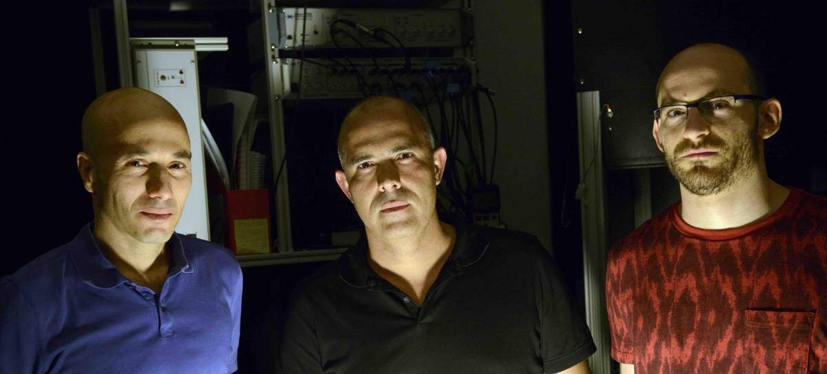
Prof. Alon Chen, postdoctoral fellow Dr. Marloes Henckens, Dr. Ofer Yizhar and research student Yoav Printz – all of the Weizmann Institute of Science’s Neurobiology Department – narrowed their search for the nerve cells that regulate the anxiety response to the extended amygdala, the region generally responsible for such emotions as fear and anxiety. This area of the extended amygdala was known to be involved in the stress response. Some of its cells express receptors for a protein that is released within the brain in stressful situations.
“We knew that this specific brain region is involved in regulating the response to stressful challenges,” says Chen. “So we decided to experiment with a technique for turning neurons ‘on’ and ‘off’ in order to learn if and how particular brain cells affect the anxiety response.” The technique, called optogenetics, uses light to control neuron activity. Lab mice were genetically engineered to express a light-sensitive protein in specific nerve cells in the extended amygdala. Then, tiny optic fibers could beam blue or yellow light onto these cells to switch their activities on or off.
Comparing mice that had these cells turned either on or off, the scientists found that those with the neurons activated showed less anxiety than the mice in which the same neurons had been switched off.
To further examine the effects of these neurons, the researchers measured levels of cortisol – a stress hormone that directs many of the body’s systems to properly respond to stressful stimuli. They compared the mice that had their extended amygdala neurons switched on with the control group. The mice with the activated neurons had lower overall cortisol levels, and it took less time for cortisol levels to return to normal after a stress-inducing event. These experiments enabled the scientists, for the first time, to identify the exact location and function of the neurons within the extended amygdala that regulate the anxiety response.
Tiny optic fibers could beam blue or yellow light onto these cells to switch their activities on or off
If these neurons regulate the anxiety response, it stands to reason they might also be involved in PTSD. To unravel the connection, the researchers first exposed the mice to a traumatic event. The mice were next transferred to a new environment and then given a reminder of the traumatic event. This can produce the symptoms of PTSD in mice as well as humans. Right after the reminder, some of the mice had the newly discovered nerve cells switched on through the optogenetic fibers. A week later, the mice were tested for PTSD. In the control group, in which the cells had been left alone, some 42% of the mice had developed the symptoms of PTSD. Only 8% of those that had received the optogenetic “turn-on” showed signs of the disorder.
“’Switching on’ these particular neurons improved the mice’s ability to recover from traumatic experience, and thus to cope with the symptoms of PTSD,” says Chen. “Locating the exact neurons and receptors involved could be crucial. The better we understand the brain’s mechanism for regulating the response to stress, the more it may help us to develop drugs that are more targeted, and hopefully more effective, for treating anxiety-related disorders.”
Prof. Alon Chen's research is supported by the Henry Chanoch Krenter Institute for Biomedical Imaging and Genomics; the Perlman Family Foundation, Founded by Louis L. and Anita M. Perlman; the Irving Bieber, M.D. and Toby Bieber, M.D. Memorial Research Fund; the Adelis Foundation; the Appleton Family Trust; and Mr. and Mrs. Bruno Licht, Brazil. His research is also supported by the Ruhman Family Laboratory for Research in the Neurobiology of Stress.