Are you a journalist? Please sign up here for our press releases
Subscribe to our monthly newsletter:
Are you a journalist? Please sign up here for our press releases

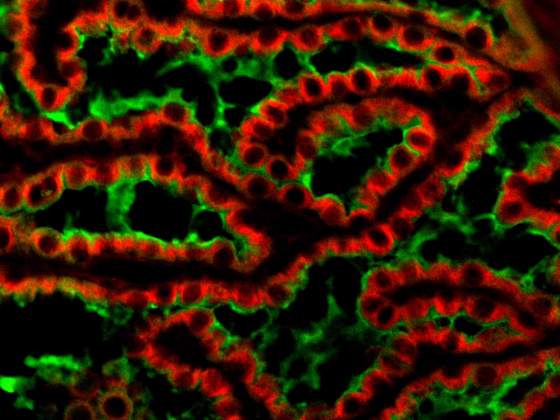
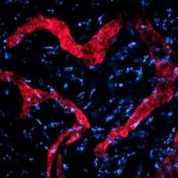
Kuti Baruch, Aleksandra Deczkowska from the Lab of Prof. Michal Schwartz in the Department of Neurobiology
The choroid plexus (in green; stained for the tight-junction molecule claudin-1) of an aged (22 months old) mouse overexpressing the arginase-1 (in red). 2013
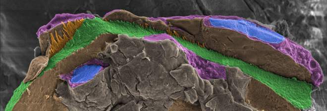
Anat Akiva from the labs of Profs Lia Addadi and Steve Weiner in the Department of Structural Biology.
Confocal fluorescence image of a zebrafish larva tail, taken with Guy Malkinson.

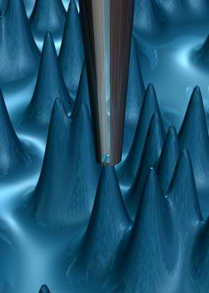
Aviram Uri from the lab of Prof. Eli Zeldov in the Condensed Matter Physics Department
SEM (scanning electron microscope) image of a part of a Multi SQUID on tip (SOT).
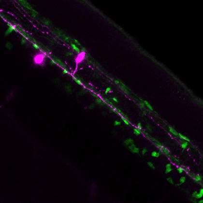
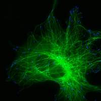
Nataliya Borodovsky from the lab of Prof. Elior Peles in the Department of Molecular Cell Biology.
Myelinated nerves in Zebrafish embryos. Spinal cord single cord neurons (in pink). The axon is myelinated with myelin sheet produced by oligodendrocytes (in green).
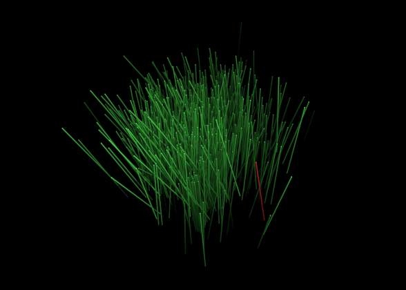
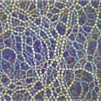
Christoffer Norn from the lab of Dr. Sarel Fleishman of the Biological Chemistry Department.
Each of the thousand blades of 'grass' represents the position and orientation of one antibody subunit and required about two years of nurturing by a crystallographer to 'grow'.
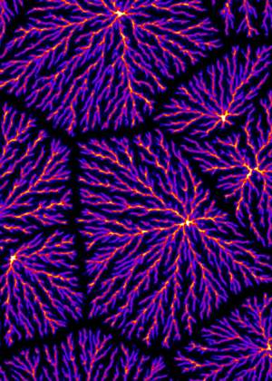
Dan Bracha from the lab of Prof. Roy Bar-Ziv in the Department of Materials and Interfaces.
A DNA brush under a condensed dendritic state with thousands of end-tethered chains in each pixel. The image was taken using fluorescence microscopy.
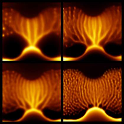
Lior Embon from the lab of Prof Eli Zeldov in the Department of Condensed Matter Physics.
Magnetic vortices in a super-conducting lead film imaged by the scanning SQUID-on-tip microscope.
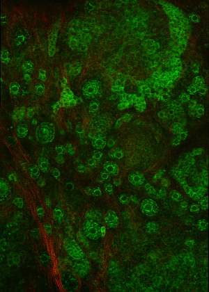
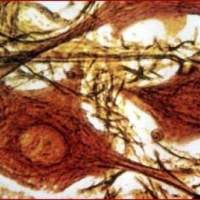
Filip Bochner from the lab of Prof. Michal Neeman in the Department of Biological regulation.
Early stages of follicle maturation in the cortex of intact murine ovary. Primordial follicles (round green structures), embedded in the collagen network (red) are at the beginning of their journey to become mature oocytes.

Oren Forkosh from the lab of Lab of Prof. Alon Chen in the Department of Neurobiology.
The behaviors shown in these panels range from exploration of the environment by a single mouse to interactions between pairs, to complex joint behavior of the whole group.
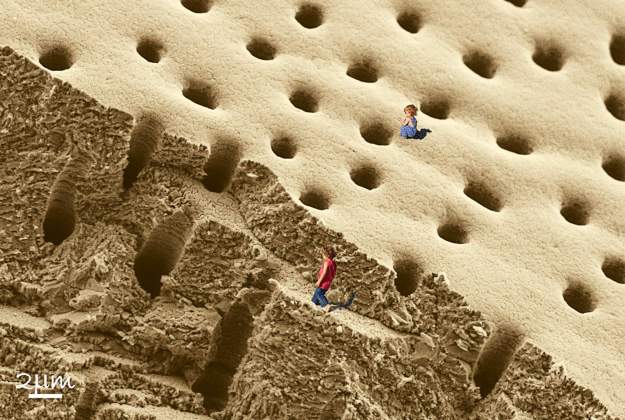
Gal Mor-Khalifa from the labs of Prof. Lia Addadi and Prof. Steve Weiner in the Department of Structural Biology.
SEM micrograph of an untreated fracture surface of the foraminifer lessonii calcitic shell wall.
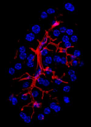
Erez Geron from the lab of Prof. Benny Shilo in the Department of Molecular Genetics.
The functional unit of the exocrine pancreas, the acinus (“grapes” in Latin), is a cluster of epithelial cells which forms and shares a joint tube. This beautiful picture was taken in an experiment gone wrong, yet I feel lucky that I imaged it.
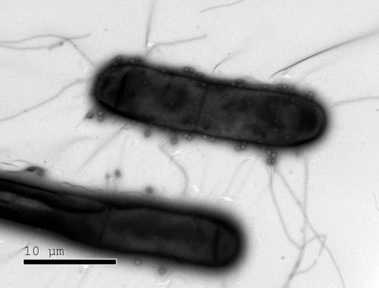
Rotem Sorek - Department of Molecular Genetics
Bacteria like these have numerous defenses against the phages (dots) that infect them.
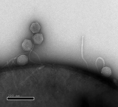
Rotem Sorek - Department of Molecular Genetics
Close up: Phages infecting this E. coli in the lab of Prof. Rotem Sorek find bacterial defense systems in place.
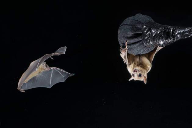
Nachum Ulanovsky - Department of Neurobiology
These bats in the neurobiology lab of Prof. Nachum Ulanovsky are helping decipher how we map the movements of others.

Neta Regev-Rudzki - Department of Biomolecular Sciences
Nanovesicles released from red blood cells infected by Plasmodium falciparum, viewed under an electron microscope. Scale bar: 100 nm.
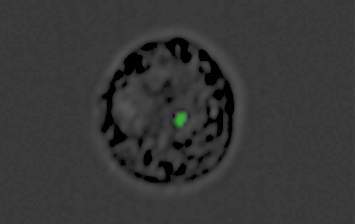
Neta Regev-Rudzki - Department of Biomolecular Sciences
A monocyte converted into a decoy by the malaria parasite: The green dot is the genetic material “cargo” inside the nanovesicle produced by the parasiteץ
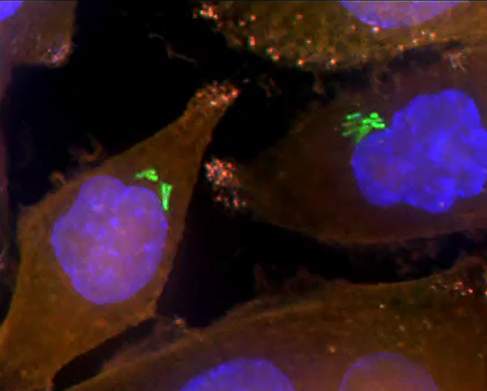
Ravid Straussman - Department of Molecular Cell Biology
Bacteria (green) inside human pancreatic cancer cells (AsPC-1 cells). The cells’ nuclei are stained blue while their cytoplasm is stained orange.
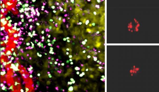
Guy Shakhar - Department of Immunology
Cancerous tumor tissue under a microscope: T cells grown under low oxygen conditions (green) and regular T cells (purple) show similar distribution patterns vis-à-vis blood vessels (red). Right: The content of granzyme B, a cell-killing enzyme (red), is much higher in T cells grown under low oxygen conditions (top) than in regular T cells (bottom).
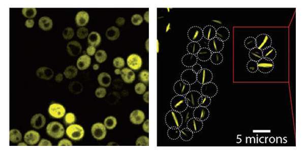
Emmanuel Levy - Department of Structural Biology
Yeast cells producing a bacterial symmetric protein complex with eight units. When it is not mutated (left), the complex diffuses freely inside the cell, but a single mutation (right) triggers its assembly into long filaments
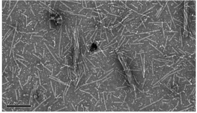
Emmanuel Levy - Department of Structural Biology
Electron microscopy of the filaments reveal Lego-like stacking of protein complexes.
Shalev Itzkovitz - Department of Molecular Cell Biology
Single-celled lining of the intestines under a microscope. Messenger RNA molecules of two different genes (red and green) are located on different sides of the cell nuclei (blue)
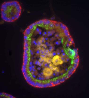
Shalev Itzkovitz - Department of Molecular Cell Biology
A circular organoid, about 0.5 mm in diameter, that mimics a cross-section of the gut in the test tube. In its outer layer, messenger RNA molecules of two different genes (red and green) are located on different sides of the cell nuclei (blue)
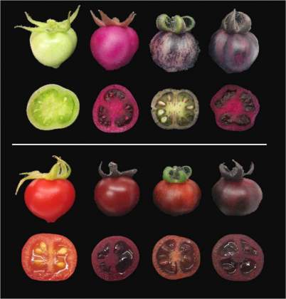
Asaph Aharoni - Department of Plant and Environmental Sciences
Unripe (top) and ripe (bottom) tomatoes. Regular tomatoes (far left) start out green and turn red when ripe. In contrast, genetically engineered tomatoes assume different shades of red-violet, depending on whether they produce betalains (second from left), pigments called anthocyanins (second from right) or betalains together with anthocyanins (far right)
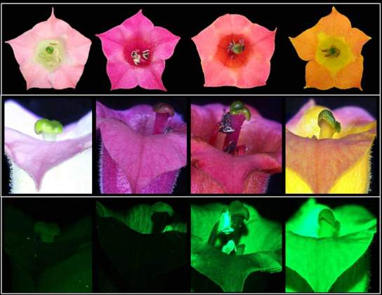
Asaph Aharoni - Department of Plant and Environmental Sciences
Tobacco flowers in nature are pale pink (far left), but can take on new colors (three images on the right) when genetically engineered to produce betalains
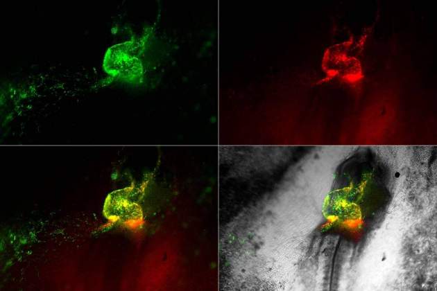
Eldad Tzahor - Department Cell Biology
The early heart tube of a chick embryo: Cardiac and endothelial cells are made visible by specifically expressing fluorescent proteins under the control of the Nkx2.5 (green) and Isl1 (red) cardiovascular genes
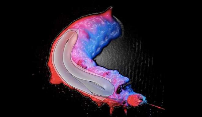
Ulyana Shimanovich - Department of Materials and Interfaces
A silkworm viewed with an infrared camera. The pale elongated cavity is the silk gland. © 2017 Natural Materials Group
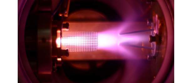
Edvardas Narevicius - Department of Chemical and Biological Physics
Cold beam experiment reveals shockwaves from the skimmer lip interfering with the beam
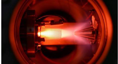
Edvardas Narevicius - Department of Chemical and Biological Physics
As the skimmer temperature is lowered, the density of the beam rises. Pulsed discharged enabled the researchers to visualize the beam density
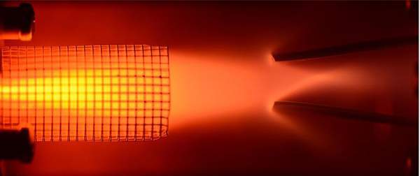
Edvardas Narevicius - Department of Chemical and Biological Physics
Skimmer experiment
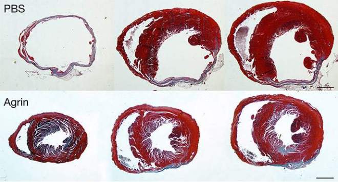
Eldad Tzahor - Department of Molecular Cell Biology
Fibrotic staining (blue) of control (PBS) and Agrin-treated hearts 35 days following myocardial infarction shows a reduction in fibrosis following treatment
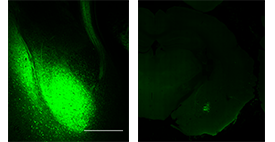
Ofer Yizhar - Department of Neurobiology
(l) The amygdala (bright green), the brain region that plays a central role in controlling emotions, sends neuronal extensions (the “cord” leading upwards) to the prefrontal cortex. (r) A highly specific set of neurons in the amygdala (bright green) that connect it with the prefrontal cortex. Viewed under a fluorescence microscope
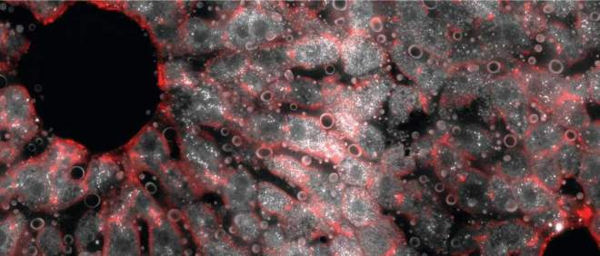
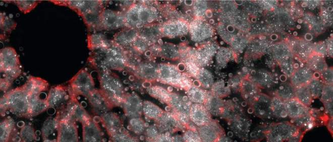
Shalev Itzkovitz - Department of Molecular Cell Biology
A cross section of a mouse liver lobule under a fluorescence microscope. The middle layer reveals an abundance of messenger RNA molecules (white dots) for the gene encoding hepcidin, the iron-regulating hormone
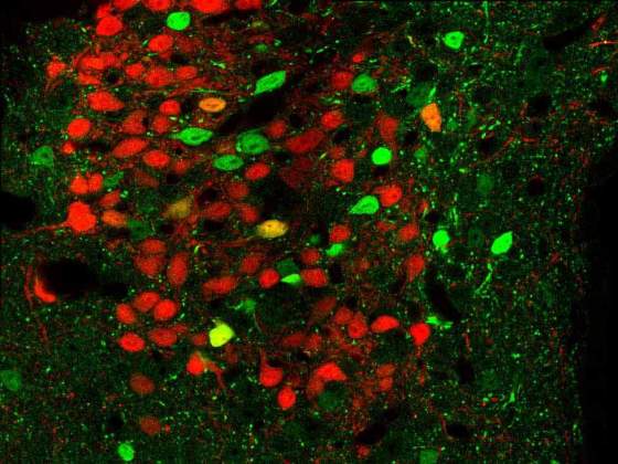
Alon Chen - Department of Neurobiology
Brain tissue from genetically engineered mice. Neurons that express CRFR1 appear in green and those that release the neurotransmitter CRF are in red. The image was obtained with fluorescence microscopy
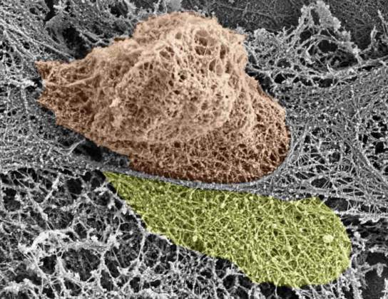
Ronen Alon - Department of Immunology
White blood cell squeezing through endothelial cells (grey) on its way out of the blood vessel walls. The actin cytoskeleton of both cells is exposed; the white blood cell nucleus is shown in brown and the large actin rich extension that dismantles the endothelial actin is shown in yellow

Avishay Gal-Yam - Department of Particle Physics and Astrophysics
In just the right conditions, the destruction of a star in a black hole's gravitational tide should produce an unusual flash of light
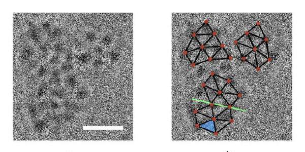
Ronny Neumann - Department of Organic Chemistry
Correlation between cryo-transmission electron microscope (TEM) images and the crystal structure. (l) TEM image showing three colliding clusters. The scale bar is 10 nm. (r) Relative positions of molecules derived from the X-ray diffraction crystal structure are overlaid (brown) on the TEM image. A twinning plane is shown (green line)
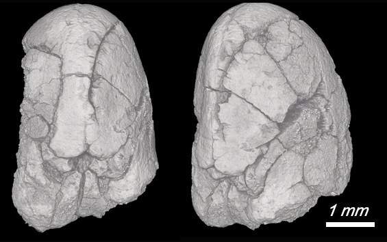
Elisabetta Boaretto - Scientific Archeology Unit , Dean for Educational Activities
14,000-year-old faba seeds contain clues to the timing of the plants' domestication
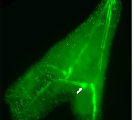
Lia Addadi, Steve Weiner - Department of Structural Biology
The green fluorescent label maps the distribution of calcium in sea urchin larvae. The label shows the elongated mineralized spicules and vesicles with large amounts of calcium
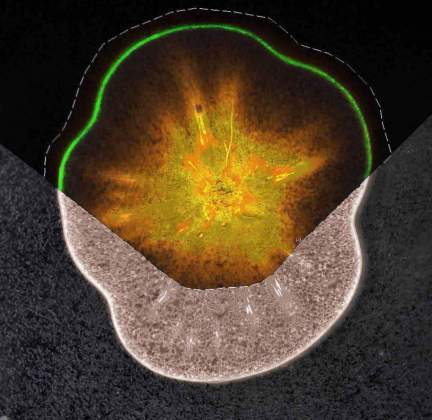
Asaph Aharoni - Department of Plant and Environmental Sciences
A section of a ripe tomato sample showing the distribution of sucrose (orange) in the flesh and of an antioxidant (green) in the fruit skin tissue; mass spectrometry imaging (MSI) technology was used to map the molecules
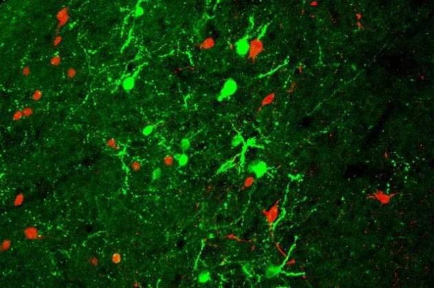
Alon Chen - Department of Neurobiology
Stress-coping molecule Urocortin-3 (green) and its receptor, CRFR2 (red), expressed in the mouse brain region responsible for social behavior. Viewed under a confocal microscope
Gilad Haran - Department of Chemical and Biological Physics
A bowtie-shaped nanoparticle made of silver with a trapped semiconductor quantum dot (indicated by the red arrow)
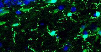
Ido Amit - Department of Immunology, Michal Schwartz - Department of Neurobiology
Microglia (bright green) in an adult mouse brain, viewed under a fluorescent microscope
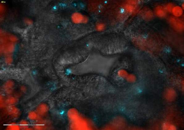
Assaf Vardi - Department of Plant and Environmental Sciences
The mouth of a coral polyp (center): Symbiotic algae are labeled in red, pathogenic bacteria that enter through this region are labeled in blue
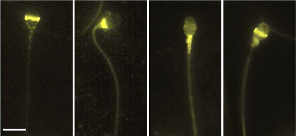
Michael Eisenbach - Department of Biomolecular Sciences
Locations of different opsins on the human sperm, viewed under a microscope, are revealed by labeling with a fluorescent antibody (bright yellow)
Yair Reisner - Department of Immunology
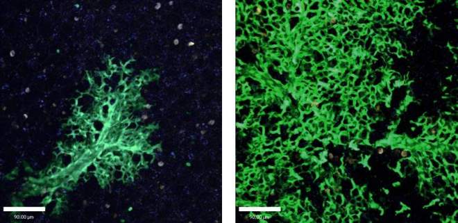
New lung cells are continuously created to replace the damaged ones: Lung tissue six weeks after stem cell transplantation (left) and 16 weeks after transplantation (right). Cells that originated in the transplanted stem cells are green, as opposed to the uncolored host lung cells. Photon-2 microscope image
Maya Schuldiner - Department of Molecular Genetics
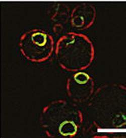
yeast proteins
Our website uses cookies to enhance user experience by remembering your preferences and analyzing website traffic.
For more information about how we use cookies please read our


