Are you a journalist? Please sign up here for our press releases
Subscribe to our monthly newsletter:

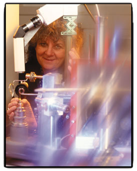
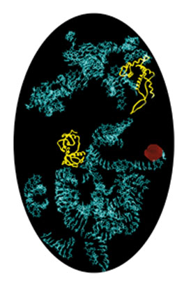
Leaning back in an office busy with mysterious images of rotating, luminescent molecules suspended in a jet-black vacuum, she laughingly recounts that her colleagues have at times called her a dreamer. Her goal, "to try to understand the principles of life from the inside by unraveling the detailed structure of ribosomes," has taken Prof. Ada Yonath of Weizmann's Structural Biology Department on a long uphill road strewn with technological and conceptual barriers.
Ribosomes are essential to life. Upon receiving genetically encoded instructions from the cell nucleus, the ribosomal factory churns out proteins - the body's primary component and the basis of all enzymatic reactions. Understanding protein biosynthesis is therefore the gateway to grasping life itself, and its darker side - the emergence of disease when production goes haywire.
This explains why ribosomes have been the target of biochemical, biophysical, and genetic studies. However, throughout nearly four decades of research, these pivotal biological units have resisted scientific attempts to reveal their functional design. To examine microscopic structures, scientists expose crystals of the material in question to high-intensity X-ray beams, a method known as X-ray crystallography. Yet the ribosome represents a daunting crystallographic challenge. Notoriously unstable, this giant protein complex also lacks the internal symmetry and repetitions that have eased the way to understanding the structure of other biological entities, such as viruses.
Indeed, back in the late 1970s when Yonath began her research, most scientists viewed her quest as a mission impossible. "Top teams around the world, such as those at UCLA and MIT in the United States and the Medical Research Council in England, had been trying to crystallize ribosomes since the 1960s - with no success. I thought, this is such a delightful group of "unsuccessful" people - they were all Nobel prizewinners - I would like to be among them," Yonath recalls, smiling.
And her determination paid off. In 1980, having tried over 25,000 conditions for growing ribosomal crystals, Yonath, who divides her time between Weizmann and the Max Planck Research Unit for Ribosomal Structure in Hamburg and Berlin, produced the first ribosomal crystal ever.
Now she has reached another scientific landmark: she has determined the structure of the small ribosomal subunit of the bacterium Thermus thermophilus at the highest resolution ever achieved. Her study recently appeared in the Proceedings of the National Academy of Sciences (PNAS).
The uniqueness of Yonath's approach lies in phasing - designing heavy atoms as markers that stand out like flares in the ribosomal map due to their high electron density. These markers significantly enhance the ability to pinpoint functional units within the ribosome.
Ribosomes consist of two independent subunits of unequal size. Yonath set her sights on 30S, the smaller subunit. More specifically, she wanted to capture "snapshots" of 30S in its active form - during the precise moment that protein biosynthesis begins. To do this, her team introduced an analogue of messenger RNA - a molecular go-between arriving from the nucleus. "The messenger attaches itself to a specific site thereby opening the gate to protein biosynthesis, which is essentially kept under lock and key," Yonath explains. Once activated and bound, it was possible to catch the 30S subunit in the act, by flash freezing the crystals at cryo-temperature (-185C or -365F).
Yonath's findings are the result of almost twenty years of experimentation. Along the way, she became the first scientist to create ribosomal crystals that diffract to high resolution, around 3 angstrom (1A = 39.37-10 inches). "My belief in the possibility of obtaining these crystals was partially inspired by natural phenomena," she says. "In nature, stressful biological conditions trigger "packing" of ribosomes into condensed crystalline structures that help prevent ribosomal damage. It is this structure that enables bears to resume protein production in the spring, following hibernation. The same thing happens in fertilized chicken and lizard eggs when shock-cooled, and in the brain cells of people suffering from dementia." Ironically, Yonath's quest for durable crystals to explore protein biosynthesis, fundamental to life, eventually led to the shores of the Dead Sea. There, two hardy strains of thermophilic (heat-loving) and halophilic (salt-loving), bacteria proved ideal candidates. "After all, they've been around almost unchanged for five million years," she explains. Yonath also pioneered cryo-cystallography, today a standard research procedure in structural biology. This approach is based on exposing crystals to cryo-temperature during X-ray measurement to minimize their disintegration.
The goal of near-atomic resolution of ribosomal structures is closer than ever before, following Yonath's latest achievements. Her approach and procedures have been repeated by a growing contingent of international researchers - all racing to unravel the mystery of ribosomal functioning. Superior strategies for fighting the pathogenic protein biosynthesis characterizing cancer cells, and improved antibiotics that target bacterial agents at the ribosomal level might yet be among the legacies of these hard-won ribosomal revelations.
Prof. Ada Yonath holds the Martin S. Kimmel Chair. Her research is supported by the Helen and Milton Kimmelman Foundation, New York.
