Are you a journalist? Please sign up here for our press releases
Subscribe to our monthly newsletter:
Each of our individual cells has its precise place and function and is able to integrate seamlessly with other various cell types to perform the tasks necessary for the body’s correct development and functionality. This fascinating process has drawn the attention of many scientists who are interested in characterizing it throughout all stages of life, as well as in sickness and in health.
To understand how cells acquire their initial specialized identity and organization, most studies focus on the earliest stages of embryonic development – a process called “gastrulation” – during which new cell types form at an astounding rate. Ensembles of individual cells adjust to their intended roles while dividing and multiplying into hundreds, then thousands, and ultimately millions of cells, giving rise to the basic scaffolding of the body’s blueprint and laying the foundation for the development of all organs and systems.
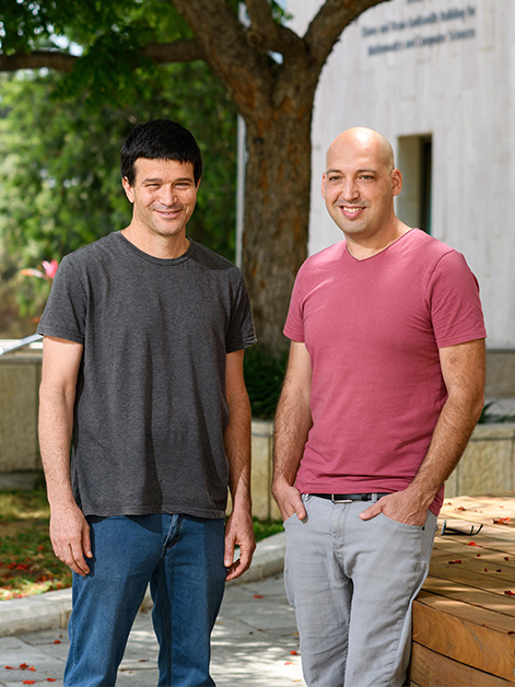
 Prof. Amos Tanay, Dr. Yoav Mayshar, Dr. Markus Mittnenzweig and Dr. Yonatan Stelzer. Likening gastrulation to dance")
Mature cells typically change and regenerate slowly, following cues and triggers from their environment. In contrast, embryonic gastrulation is rapid and happens in a perfectly reproducible fashion. This provides scientists with an opportunity to explore the proliferation and differentiation of cells, time and again, and to search for answers to such riddles as why some cells become a fully functional brain, while others are directed to make up our immune system. Yet, since the mammalian embryo is a tiny, fast-growing structure protected by its mother’s womb, scientists have so far only been able to study gastrulation indirectly, focusing on each of its separate aspects one at a time, rather than capturing the process as a whole.
Weizmann Institute of Science researchers Dr. Yoav Mayshar of Dr. Yonatan Stelzer’s group at the Molecular Cell Biology Department, and Dr. Markus Mittnenzweig of Prof. Amos Tanay’s group at the Computer Science and Applied Mathematics Department and the Biological Regulation Department, together with colleagues from both groups, have devised a new method of tracing the intricate process of gastrulation in a mouse model by employing state-of-the-art single-cell genetic analysis technologies and advanced mathematical modeling. Many scientists around the world have contributed to creating detailed atlases of cell types from numerous organisms. Still, the team at Weizmann believed that a deeper understanding of differentiating embryonic cells and their dynamics calls for a fresh approach.
The result is a “moving picture,” which is composed of discrete cellular “frames,” describing how embryonic cells change together over time
Single-cell genetic analysis usually affects the cell’s proper functioning, oftentimes leaving researchers with a lot of guesswork as to how exactly cells develop and differentiate. Thus, for example, it was assumed that cells take the shortest route from their baseline activity to full differentiation. “If we should liken gastrulation to dance,” says Mittnenzweig, “then in our former models we assumed that each dancer, or cell, is a soloist looking for the shortest way between entering the stage and exiting it.” However, Mayshar adds, “This does not comply with developmental biology as we understand it today, following many years of experimentation.”
In the new study, the researchers created a unique combination of genetic data obtained from thousands of single cells and physical measurements of whole embryos. This enabled the reconstruction of the developmental process by identifying minute changes in embryonic morphology, a common practice in developmental biology, together with mathematical modeling that takes into account the gene expression profiles of cells in each separate embryo. The result is a “moving picture,” which is composed of discrete cellular “frames,” describing how embryonic cells change together over time.

The researchers noticed that while some cells, such as those involved in the development of essential organs, immediately hone in on their target, others uphold a fairly broad developmental potential. This strengthens the hypothesis that cells differentiate through a dynamic process of continuous transformation and that they generally possess immense potential for such transformation not only in the earliest but also in the later stages of their existence. The potential for transformation of these cells exists as part of a composite structure where each cell contributes its part to this dynamic multicellular “ecosystem.” In this setting cells engage in give and take with their neighbors while the transcriptional landscapes of individual cells adjust to a constantly changing environment – both temporally and spatially.
Returning to our previous dance simile, we can now say that this model, in a way, likens gastrulation to a virtuoso dance number where each dancer, or individual cell, moves in an unknown trajectory but often in unison with other cells. Together, and in perfect harmony, they form orchestrated ensembles that ultimately develop into functioning tissues and organs, a choreography that accurately repeats itself during the initiation of life.
The new model for studying gastrulation offers an advanced tool kit that makes this intricate process more approachable, thus allowing researchers to better track cells as they develop and differentiate. Addressing questions such as why and how cells take on their specific roles within the tissue, and how they affect their environment and are affected by it in return could improve scientists’ understanding as to why this outstanding choreography sometimes goes awry, distressing the organism’s health and ultimately risking its survival.

During only 2 days of gastrulation the mouse embryo grows 64-times its initial size: If it would have continued to grow at this rate, at birth its weight would amount to over 50 kilograms instead of its actual 2 grams.
Dr. Yonatan Stelzer is the incumbent of the Louis and Ida Rich Career Development Chair.
Dr. Stelzer's research is supported by the Helen and Martin Kimmel Institute for Stem Cell Research; the Moross Integrated Cancer Center; the Hadar Impact Fund; Janet and Steven Anixter, JoAnne Silva and the Lester & Edward Anixter Family; the Maurice and Vivienne Wohl Biology Endowment; Barry and Janet Lang; the Estate of Emile Mimran; and the European Research Council.
Prof. Amos Tanay's research is supported by the Ilana and Pascal Mantoux Institute for Bioinformatics; the Wolfson Family Charitable Trust and Wolfson Foundation; and Barry and Janet Lang.