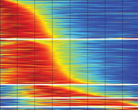
What's the difference between cancer cells that are killed by chemotherapy and the ones that survive the treatment? To tackle this question, Ariel Cohen together with Naama Geva-Zatorsky and Eran Eden in the lab of Prof. Uri Alon of the Molecular Cell Biology Department developed an original method for imaging thousands of living cells and analyzing their activities automatically by computer.
The team members worked for several years to complete the project, which entailed observing the behavior of over 1,000 different proteins, tagging specific proteins in each group of cancer cells and each time capturing a series of time-lapsed images over 72 hours. A chemotherapy drug was introduced 24 hours into this period, after which the cells began the process of either dying or defending themselves against the drug.
The team's efforts have produced a comprehensive library of tagged cells, images and data on cancer cell proteins – a virtual gold-mine for further cancer research. And they succeeded in identifying two proteins that seem to play a role in cancer cell survival. One of them, known by the letters DDX5, is a multi-tasking protein that, among other things, plays a role in initiating the production of other proteins. The other, RFC1, also plays a variety of roles, including directing the repair of damaged DNA. When the researchers blocked the production of these proteins in the cancer cells, the drug became much more efficient at wiping out the growth. Says Cohen: "We were able to pinpoint possible new drug targets and to see how certain activities might boost the effectiveness of current drugs."
Prof. Uri Alon's research is supported by the Kahn Foundation; Keren Isra - Pa'amei Tikva; and the Minerva Junior Research Group on Biological Computation.
