Biological materials and systems are multifunctional, adaptive, nonlinear, complex, and, in general, just "weird and wonderful.”
- Biologist Stephen Wainwright, Duke University
In the early 1940s, Swiss engineer Georges de Mestral went for a walk in the woods with his dog. Upon his return he noticed they were both covered with seeds of a burdock plant.
Removing the burs proved a frustrating task. Taking a closer look, de Mestral observed that the burs had hundreds of tiny little hooks - a clever trick for a plant wishing to disperse its seeds. He thought this might be an ideal means of joining two fabrics together - and the idea of Velcro was born.
Today Velcro is found on everything from zippers to sneakers to space suits, and scientists are working on yet another form that rips apart silently - a critical ingredient for military operations.
Velcro is a classic example in the story of biomimetics, a field that has scientists turning to nature to examine its countless bioengineering feats, polished over time. The hope is that new ideas for advances in medicine, technology and industry might be hidden in some of the solutions evolved to meet the challenges of life - from bones and teeth, capable of withstanding decades of grinding wear and tear, to the enormous tensile strength and elasticity of spider dragline silk, which ounce for ounce is stronger than steel, enabling a dangling spider to safely dive down and nab its prey.
Dealing with the daily grind
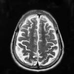
Birds don’t have ‘em, nor do bees; sharks can replace them almost immediately if they break; and we humans have teeth that, excluding sugar junkies and night grinders, can last a lifetime given the right dental care.
A team of Institute researchers has now discovered a key factor explaining this hardiness: a spring-like structure that absorbs the mechanical stress incurred as teeth chew, grind or, as circumstance would have it, chatter their way through an arctic freeze. Interestingly, this structural feature constitutes only a tiny percentage of the overall tooth.
The team, consisting of Prof. Stephen Weiner of the Structural Biology Department and post-doc Dr. Rhizi Wang, began by scouring the literature in search of a dental component that might fit the bill of mechanical stress absorber. “Drawing on his materials science expertise, Wang put his finger on the core question,” says Weiner. “Given that the two main components of teeth are enamel - a hard, brittle material - and dentin, which is much softer, Wang hypothesized that there must be an intervening layer that allows these two very different materials to work together.”
The researchers knew that studies dating back to the 1960s had uncovered a certain structural zone positioned directly between enamel and dentin, but they found no follow-up studies exploring its significance. They decided to determine whether teeth actually compress preferentially in this unexplored area when subjected to mechanical stress.
As published in the Journal of Biomechanics, the team found that the structure in question, which they dubbed the “soft zone,” is where almost all compression takes place. The study showed that the zone - which is merely 200 microns thick, compared to enamel’s 1,000 microns and dentin’s 5,000 - is in fact the “working part” of the tooth. When we chew, the enamel part of our teeth is pushed back while the soft zone compresses. It then functions like a spring, bouncing back after the compression.
Together with Prof. Asher Friesem and dentist Paul Zaslansky, now a Ph.D. student at the Institute, the team is currently using a new technique known as speckle interferometry to map the deformation of objects in response to an applied force, at a resolution of tens of nanometers. An enhanced understanding of dental stress absorption mechanisms should prove valuable in tooth reconstruction.
Biological computer diagnoses cancer - in a test tube
Trillions of microscopic computers vigilantly patrolling your body in search of disease? Institute researchers have recently made a pioneering step in this direction, developing a biological computer able to identify - in a test tube - molecular changes indicative of certain cancers, to diagnose the type of cancer, and to react by producing an appropriate drug.
The study, recently published in Nature, was performed by Prof. Ehud Shapiro of the Departments of Computer Sciences and Applied Mathematics and Biological Chemistry, his research students Yaakov Benenson, Binyamin Gil and Uri Ben-Dor, and Dr. Rivka Adar.
The team programmed their computer to detect prostate cancer and one form of lung cancer. The computer evaluates four genes that become either under-or overactive once the disease sets in. The genes chosen control the expression of messenger-RNA (which carries information from the nucleus to the ribosome, the cell’s protein factory). The scientists introduced different levels of these RNA molecules into the test tube to simulate the presence or absence of cancer.
Made entirely of biological molecules, the computer has three components - input, computation and output. The first consists of short strands of DNA, called transition molecules, which check for the presence of the messenger RNA produced by each of the four cancerous genes.
The second component is a computation (diagnostic) module, consisting of a hairpin-shaped long DNA strand. As the computer’s input component checks for the presence or absence of the four cancerous markers, this diagnostic unit checks each input in turn, producing a positive diagnosis of malignancy only if all four markers point to cancer.
This diagnostic module also contains the computer’s third component: a single-stranded DNA that is known to interfere with the cancer cell’s activities. In the case of a positive diagnosis, the unit releases its hold on the therapeutic unit, activating its cancer-fighting potential.
Shapiro’s team first went on record in 2001, when it created the first autonomous biological nanocomputer. Made entirely of biological molecules, the computer's input, output, and “software” were made up of DNA molecules. The device was so small that as many as a trillion such computing devices could work in parallel in one drop of water, collectively performing a billion operations per second with greater than 99.8 percent accuracy per operation. It was recently awarded the Guinness World Record for the “smallest biological computing device.”
Shapiro: “Our study offers a vision of the future of medicine. It is clear however, that it may take decades before such a system operating inside the human body becomes a reality.”
Computers in the dust
In 1954 researchers at the Weizmann Institute built the first electronic computer in Israel, and one of the first computers worldwide, which they fondly named WEIZAC. Thirty years later, silicon chip computers routinely functioned at a rate that was thousands and millions of times faster than WEIZAC.
Today, research at the Institute targets the development of ever-faster, more compact chips designed according to the emerging principles of quantum electronics. These will inevitably leave silicon chips in the dust, much as the silicon chips once turned WEIZAC into a museum exhibit.
Yet even before these quantum electronic chips have become a reality, they already have a potential successor - the tiny biological computer developed by Prof. Shapiro’s team.
The slick joint
Body joints are superbly lubricated. They have to be. As the meeting point between every two bones in our body, they are what makes our every movement possible - from walking, bending and maneuvering our fingers to playing ball and dancing. And they’re supposed to last a lifetime.
Mimicking key design elements of this biolubrication system, physicist Prof. Jacob Klein of the Weizmann Institute of Science, has recently created a synthetic lubricant that cuts friction by a thousand-fold or more. The study, published in Nature, could lead to a range of applications - from longer-lasting micromachines and higher-density hard disks to biomedical devices, including improved hip transplants and treatment for dry eye syndrome.
Previous studies had suggested that biolubrication systems, including those in our joints and eyes, may contain hyaluronan, a molecule that coats the rubbing surfaces of our joints, shielding them from mechanical damage. Hyaluronan was also known to be strongly attracted to water. But how these two factors combined to create the most effective lubricatory system anywhere, remained a mystery.
Klein and colleagues suspected that in joints, hyaluronan may be attached to a thin cartilage layer covering the bone, while parts of this long, chain-like molecule stick out into the synovial fluid between the bones, similar to bristles on a hairbrush. These bristles or others playing a similar role, they believed, function as the body’s lubricants.
To test their theory, the team developed a synthetic model that mimicked a double-brush system, anchoring two charged molecules (polyelectrolytes) to opposite-facing ceramic surfaces. The resulting system showed extremely effective friction resistance, particularly when exposed to a water-based solution. “The brushes strongly try to avoid each other, resisting penetration even when an external force is applied that presses them closer. This enables them to easily slide past one another,” says Klein.
The superior lubrication may largely be explained by electrical charges. The synthetic brushes were designed to imitate the electrically-charged nature of biolubricants. Similar to the negative charge characterizing the hyaluronan molecule, for instance, the end of the synthetic brushes stretching away from the ceramic surface was designed to be negatively charged. This negative charge then attracted water molecules - which in fact explains why the brushes performed most effectively in a water-based solution. “The water molecules are tightly bound to charges on the brushes, causing them to act like molecular ball bearings to reduce friction,” says Klein.
Other players in this charge cast are the small mobile ions trapped in the space between the bristles. According to Klein, when the brushes approach one another, their respective clouds of positive ions are mutually repellant, increasing the brushes “distaste” of penetrating one another.
“Our general idea was to draw on nature, says Klein. “It made sense to try out the design principles evolved and optimized over millions of years.”
Humpty-Dumpty mechanics
Two hundred and six - this is the number of bones in the adult human skeleton. Over the years of your life, as you walk, play basketball, dance through the night or bungee jump from a steep cliff, these bones will see you through, supporting your weight when you stand upright and protecting your brain and internal organs.
How bones withstand the immense array of mechanical stresses imposed upon them has long fascinated scientists, who have set about probing their chemical and biological properties and even crushing them to determine the relationship between their structure and their outstanding performance. Yet despite several decades of research, a full understanding of this complex biological material remains elusive.
The beauty of bone stems from two key properties - it is a composite material and it is highly hierarchical in structure. The principle of forming a composite is to take two materials, often highly different from each other - say, a soft and an extremely hard material - weave them together somehow, and, presto, a new material is born that is generally far superior to either of its individual components.
In bone, this partnership comes in the form of a ceramic-like, brittle mineral salt called hydroxyapatite, which is bolstered by collagen (a protein) that adds elasticity and strength. The tissue has several levels of organization, ranging in scale from nanometers to centimeters, each with a different structure and function. Adding to this complexity is the fact that bone changes over time, with specialized bone cells actively replacing older bone tissue in a process that has adults renewing their entire skeleton roughly every 7-10 years.
In the 1990s Prof. Stephen Weiner of the Institute’s Structural Biology Department and Prof. Daniel Wagner of the Materials and Interfaces Department collected data on the micro-mechanical properties of bone and developed a mathematical model explaining how bone’s various components affect its mechanical function, including bone elasticity. The model made it possible to predict the mechanical properties of bone with unprecedented accuracy.
Weiner is currently working to further understand the contribution to mechanical strength made by the individual cylinders within bone. Each of these cylinders changes over time, altering its mineral content and thus its mechanical properties. “While past studies succeeded in determining the average mechanical properties of bones, what we really need to know is how each of these individual cylinders responds to stress,” says Weiner. Insights at this structural level may yield new treatments for osteoporosis and other bone diseases.
When bones become porous
Osteoporosis, or porous bones, is a disease characterized by structural deterioration of thebone tissue, leading to low bone mass, fragility and increased vulnerability to fractures of the hip, spine, and wrists.
Generally appearing at age 50 or above, osteoporosis is caused by unbalanced bone “remodeling,” as cells responsible for breaking down old bone tissue (osteoclasts) become more active than those laying down new tissue (osteoblasts), causing bone loss.
The disease affects more than 44 million people in America alone, 68 percent of whom are women, causing an estimated annual national expenditure of $17 billion.
Fine spine choices
Back when marine animals first began trying on mineralized, exoskeleton armor, over 550 million years ago, sea urchins had a crucial “choice” to make. The odds pointed to calcite, an abundant calcium carbonate mineral, as a natural candidate for their skeletal and spine construction. But while clearly the “cheapest” material available, calcite seemed a fragile choice at best - it’s an extremely brittle material, easily shattered by mechanical stress. Some of their contemporaries opted for silica, magnetite or calcium phosphate. Yet sea urchins stuck with calcite. They then proceeded to evolve unique biological adaptations to dramatically enhance its material strength.
At the Institute, Profs. Stephen Weiner and Lia Addadi have discovered key features that explain the surprising strength of sea urchins, as well as of mollusks and bones - all natural composites.
Scientists had long been unable to explain the durability of sea urchin spines, which they believed consisted entirely of a single calcite (and thus highly fragile) crystal. The Weizmann scientists revealed that while calcite indeed constitutes 99.9 percent of the urchin spine, the remaining 0.1 percent consists of proteins entrapped within the crystalline matrix. And amazingly, that is what makes all the difference.
Working with researchers from the Brookhaven National Laboratory in New York, the Weizmann team discovered how the entrapped proteins act to reinforce urchin calcite against fracture, preventing cracks from spreading through the crystal. The scientists grew pure calcite crystals in solutions containing proteins extracted from urchin spines. They found that the proteins integrated into the crystal, altering its structure and making it far less brittle. The question remained, however, how a component present in such minuscule proportions could have such far-reaching effects on material quality.
Follow-up studies showed that the proteins had integrated into the pure crystals preferentially, along planes that are oblique to the crystal cleavage plane, thus disrupting fracture propagation. “The crystal's ability to withstand mechanical stress is enhanced because resulting cracks do not rip catastrophically along the cleavage planes but are diverted along the protein-induced planes, which absorb the force of impact,” explains Weiner.
These findings might lead to the development of new crystal-polymer composites for building lightweight, tougher ceramics and improved electrooptic materials.
Composite cornucopia
You shall no longer give the people straw to make brick as before (Exodus 5:6-7).
This is the first recorded account of a man-made composite, describing bricks used in ancient Egypt, which were made of mud mixed with straw. The straw increased the bricks’ thermal stability, preventing them from cracking. Modern studies show that these bricks were three times stronger than those lacking straw. Thousands of years later, the Egyptian landscape is still dotted with monuments made of such bricks.
Composites are everywhere. Though often referring to the fiber-reinforced metals, polymers and ceramic materials originally developed for use in the 1950s, composite superstars include those found in nature - from human bone, to tree trunks, sea shells and spider dragline silk, which ounce for ounce is stronger than steel and tougher than kevlar.
“Sightless” marine creature found to be all eyes
A team of scientists has found that the brittlestar, a marine invertebrate long thought to be sightless, is in fact covered with calcite crystals that function as optical receptors for a compound eye. A better understanding of how to build such high-quality microlenses may lead to improved computer circuits.
Profs. Lia Addadi and Stephen Weiner of the Weizmann Institute’s Structural Biology Department had long been interested in the ways in which animals build their skeletal structures. When they met Dr. Gordon Hendler of the Natural History Museum of Los Angeles County, Hendler brought to their attention one particular species of brittlestar that appeared to be particularly sensitive to changes in light, quickly escaping into dark crevices at the first sign of danger. He suspected that spherical crystal structures on the brittlestar’s outer skeleton serve as lenses, transmitting light to its nervous system.
By analyzing the geometry of the crystal lenses Addadi and Weiner, together with their then graduate student Joanna Aizenberg, were able to pinpoint the expected focal point on the nerve bundles below, but they lacked the means of proving that these lenses indeed transmit light to the nervous system within.
This is where things stood for almost ten years, until the team came up with the idea of examining the lenses using lithography, a semiconductor technology.
Placing one of the crystals above a layer of photosensitive material, Aizenberg exposed the system to light and found that it reached the photosensitive tissue in spots directly underneath the crystals. Her findings also demonstrated that the crystalline lenses act as “corrective glasses,” filtering and focusing light on photoreceptors within the nerves. But unlike man’s ability to see in virtually only one direction, this complex visual system enables the brittlestar to detect approaching danger from many directions. The lenses expertly compensate for common optical distortion effects. This unique visual architecture has prompted hopes for new materials that would mimic the brittlestar model. Knowing how to build such microlenses of high optical quality could lead to improved microlithography tools used in etching the integrated circuits found in computers.