Are you a journalist? Please sign up here for our press releases
Subscribe to our monthly newsletter:

A doctoral student with a degree in physics, who conducts research on biological systems in the Faculty of Chemistry, has received an award given by the Faculty of Mathematics. To Roie Shlomovitz, a student in the group of Prof. Nir Gov of the Chemical Physics Department and the 2010 recipient of the Lee A. Segel Memorial Prize in Theoretical Biology, this makes perfect sense. Prof. Segel, a member of the Weizmann Institute’s Mathematics and Computer Science Faculty for many years who passed away in 2005, was one of the first to cross the boundaries between the disciplines of mathematics and the life sciences, showing that mathematics could be used to describe the dynamics of biological systems and teaching biologists to think “mathematically.” “My work fits right in with Segel’s approach,” says Shlomovitz.
Shlomovitz and Gov investigate proteins that form the cell’s internal skeleton. These proteins – actin and myosin – make up the fibers that enable our muscles to contract and are involved in cell movement and cell division. When a cell moves, actin filaments gather on the leading side, forming a bulge that propels the cell onward. In cell division, actin and myosin proteins create a ring encircling the cell’s midriff, squeezing the membrane tightly and pinching it in two.
What causes these proteins to push the cell membrane outward in one case and constrict it in another? How do they “know” when and where to apply pressure? To address such questions, Shlomovitz and Gov observe what happens to these proteins in cells, turn the data into mathematical models and, finally, test the predictions of their models in such biological systems as yeast. According to their model, actin and myosin proteins get their directions from the shapes of molecules in the cell’s membrane. When the cell is about to move, the membrane points the way by exaggerating its outward curve on one side. The convex membrane molecules “invite” the actin to gather round; large amounts of these molecules and the actin flock to the site to drive the edge forward. In the early stages of cell division, on the other hand, the membrane’s center takes on an inward curve. Investigating bacterial proteins, the researchers found that concave curves attract these proteins, as well: A circular network of actin and myosin marks the division line.
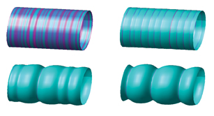
The mathematical model they developed borrowed an equation from physics that describes the free energy of a system. Using this equation, they calculated the distribution of proteins in the membrane and revealed how this distribution relies on the membrane’s shape. The researchers showed exactly how the bowed segment induces the proteins to encircle the membrane right at the center, how the growing inward bend of the membrane continuously attracts more proteins to the spot to increase the constriction, and how the distance between one protein and the next determines whether they’ll become a part of the tightening band or form separate rings.
Their model not only predicted the behavior of these proteins on artificial membrane bulges and indents; an experimental group in Taiwan found it could also explain the wavy shapes of living cell membranes. Shlomovitz: “We describe the complex biological processes occurring within a single cell by constructing chemical and physical models. Mathematics is the ‘language’ we use to analyze them.”
Prof. Nir Gov’s research is supported by the Helen and Milton A. Kimmelman Center for Biomolecular Structure and Assembly.