Are you a journalist? Please sign up here for our press releases
Subscribe to our monthly newsletter:
 Profs. Stephen Weiner and Lia Addadi and Julia Mahamid. Like shell, like bone")
In the search for new and better materials, many a scientist has turned to the study of living organisms. Generally these studies have focused on such tissues as bone or shell – complex mixtures of minerals, proteins and sugars that result in hard, strong, fracture-resistant materials. Recent research in the Institute's Structural Biology Department has shown that bones and shells share similar formation processes.
Bone and shell are composite materials in which the mineral component forms within a framework of soft macromolecules such as proteins. The framework is essentially a "mold" that directs the mineral formation. Bone grows continuously, layer by layer; but, unlike shell, it also undergoes constant remodeling. This makes it hard for scientists studying bone to distinguish new material from old, much less to trace the process by which bone is formed. Because of this difficulty, conflicting theories have arisen as to the mechanisms of bone formation. Many scientists thought that the minerals precipitate directly from a solution in the way that stalactites and stalagmites grow from elements dissolved in water. But findings published in the 1960s already hinted at the possibility that a different process was at work.
Ten years ago, research in a group headed by Profs. Lia Addadi and Stephen Weiner of the Institute's Structural Biology Department confirmed these earlier observations for shells. They showed that organisms first produce packets of amorphous material (unorganized material, as opposed to the ordered organization of crystal). These packets are transferred from the inside of cells to the building site – the spot where the mineralized tissue eventually forms. There, the packets undergo structural changes that turn them into a hard crystal. This observation triggered widespread interest in amorphous precursor mineral phases, and many different invertebrate mineralization processes were investigated.
Addadi and Weiner revealed this process in sea urchin spines, and their findings have been joined by a body of global research confirming that this method of construction is common to many different invertebrates that have shells, spines or other hard body structures. Addadi: "The original idea of precipitation from solution would require huge quantities of liquid to flow from inside the mollusk to the outside of its body. Transferring the materials in solid form – 'bricks and cement' – is a much more energy-efficient way of doing things."
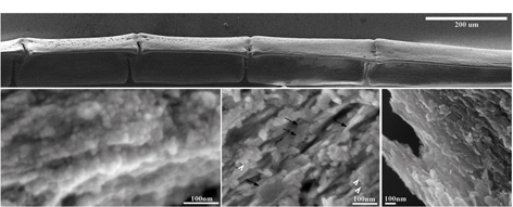
While the issue has been more or less settled for the shells of invertebrates, the question of how vertebrate bones are formed remained unresolved. This is partly because of the difficulty inherent in attempting to observe a substance with no fixed location that exists for only a fleeting stage of growth. Recently, however, research student Julia Mahamid, together with Addadi and Weiner, found a biological system that enabled them to follow bone formation step by step and identify the processes taking place at each stage. This system is the fin of a small aquarium denizen called a zebrafish. The zebrafish fin bones, which continue to grow throughout the fish's lifetime, form a sort of fan. Each bony rib of the fan is composed of segments, and the segments nearest the fin's edge are always the newest. Thus the segments can be studied as a sort of timeline of bone formation. In addition, zebrafish, which live in relatively cold water, grow slowly, and the leisurely pace of their bone development enabled the scientists to get a good look at each stage.
Using both light microscopy and scanning electron microscopy, the scientists succeeded in observing abundant spherical parcels of amorphous mineral material in the newly formed fin bone. Their findings, which appeared in the Proceedings of the National Academy of Sciences (PNAS), USA, showed that about half the mineral makeup of the newer bone segments is amorphous – a fraction that dwindles in the segments farther away from the fin edge. Their observations suggest that the amorphous material does, indeed, turn to crystal over time.
These findings are shedding light on a number of scientific mysteries, giving scientists a unique perspective on how the hard substances in our body – bones and teeth – are formed. The scientists hope that a deeper understanding of these biological processes may, in the future, help researchers find cures for diseases involving faulty bone development or repair.
Prof. Lia Addadi's research is supported by the Clore Center for Biological Physics; the Ilse Katz Institute for Material Sciences and Magnetic Resonance Research; the Helen and Martin Kimmel Center for Nanoscale Science; the Helen and Milton A. Kimmelman Center for Biomolecular Structure and Assembly; and the Carolito Stiftung. Prof. Lia Addadi is the incumbent of the Dorothy and Patrick Gorman Professorial Chair.
Prof. Stephen Weiner's research is supported by the Kekst Family Center for Medical Genetics; the Helen and Martin Kimmel Center for Archaeological Science; the Helen and Milton A. Kimmelman Center for Biomolecular Structure and Assembly; and George Schwartzman, Sarasota, FL. Prof. Weiner is the incumbent of the Dr. Walter and Dr. Trude Borchardt Professorial Chair in Structural Biology.