Are you a journalist? Please sign up here for our press releases
Subscribe to our monthly newsletter:

Having completed a working draft of the human genome, widely hailed as one of the most significant intellectual achievements of all time, one might think that those involved would head home for a well-earned vacation.
Far from it. They've rolled up their sleeves and are moving on with added speed. We now know how genes perform the cell's work, instructing it to string together different amino acids to create, say, a protein triggering blood clotting following injury, or a stomach enzyme to aid in food digestion. The next challenge is to better understand these gene products.
Proteins serve as the body's primary component and the basis of all enzymatic reactions, so the slightest change in their structure or function can lead to disease, even death. Being there -- in the right place, at the right time, and in the proper amount, is what the protein story is all about.
It's also about folding. The primary property influencing a protein's function is its 3-D structure. Consisting of one long chain, or several, the protein molecule is generally coiled or folded. Any damage to this structure can impair a protein's biological properties -- which is why, for instance, heat-wave temperatures of over 50o C can be life threatening.
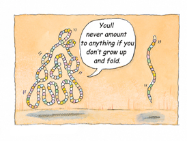
But proteins are "born" unfolded. When first produced by the ribosomal factory (which implements genetically encoded instructions arriving from the cell nucleus), proteins emerge as straight, unfurled strings. They must fold into their correct form to become functional. How does the "young" protein know how to do this? Do "folding instructions" arrive with the genetic package, or do these tabula rasa proteins receive some help from nearby friends?
Back in the 1980s Drs. John Ellis and Costa Georgopoulos (then at the University of Warwick and the University of Utah, respectively) discovered that proteins, in fact, have molecular mentors. Dubbed "protein chaperones," they walk the newborn protein through its first folding steps and provide a safe environment in which to do so. Understanding these chaperones, themselves proteins, is the main interest of Prof. Amnon Horovitz of the Weizmann Institute's Structural Biology Department. Horovitz studies the GroEL protein found in E. coli bacteria -- a molecule containing two back-to-back rings, with a cavity at each end. Folding takes place within these cavities with the help of an additional protein called GroES, which serves as a "lid," covering the cavities' exits to prevent the newborn proteins from falling out while folding.
It turns out that the chaperone molecule has two basic states. Initially, newborn proteins can easily enter the chaperone and attach themselves to its cavity walls. At this point, the chaperone molecule undergoes a dramatic structural transformation. Its cavity walls reject the newborn proteins, heaving them into the center where they can fold safely.
Horovitz: "In the past, people believed that proteins folded into their characteristic structure to attain the lowest possible energy level, just as a ball placed on a mountaintop rolls to the bottom. In other words, existing in the folded as opposed to the unfolded state requires far less energy. But what if there is a series of mountains with valleys in between? The ball might reach a relatively high valley, where it will stay put unless 'freed' by an earthquake. From a protein's perspective, this 'earthquake' can be any of a number of environmental changes, such as temperature changes or the presence of a solvent. Both can cause protein misfolding, resulting in disease." For instance, Creutzfeldt-Jakob disease, popularly known as mad cow disease, is caused by misfolded proteins that are otherwise normal.
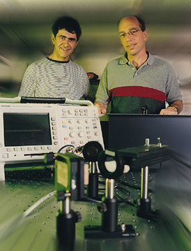
To discover how chaperones prevent protein misfolding, Prof. Horovitz is collaborating with Dr. Gilad Haran of the Chemical Physics Department. In standard experiments, billions of molecules are examined simultaneously; but Haran examines them one by one. This means that experimental results are not merely an average of different molecules, which might differ in their conformation or environment, but a representation of the full molecular variety of the process under examination.
To study the chaperone, Haran tags it with a fluorescent molecule that emits light when activated by a laser. When the chaperone protein shifts from one state to the other, it affects the fluorescent molecule's properties. In turn, a microscope equipped with sensitive light detectors picks up these changes, enabling the scientists to track the chaperone's transition cycles and obtain a better understanding of the folding processes it hosts.
But Horovitz and Haran want an even closer look. To obtain this, they plan to fasten two fluorescent molecules onto different sites of an unfolded protein. Since the wavelengths emitted by these molecules and the relative intensity of their emission are dependent on their distance from each other, the researchers believe that this approach may offer an intimate glimpse into the chaperone during protein folding.
Achieving a better understanding of how proteins fold is the next greatest challenge in what promises to be biology's century. The Human Genome Project was only the beginning -- there's still a great deal of unfurling ahead.
