Are you a journalist? Please sign up here for our press releases
Subscribe to our monthly newsletter:

Accurately detecting motion – whether that of a snake slithering though grass or a bike courier racing through a crosswalk – is vital to our survival. Yet, as the developers of autonomous vehicles have learned, identifying the trajectory of movement through space is one of the most challenging computational problems. Perceiving movement starts with cells in the retina that are anatomically built to sense a particular direction – either up or down, left or right. These roles were thought to be “built-in,” but scientists at the Weizmann Institute of Science recently showed that these so-called direction-selective ganglion cells have more flexibility than was thought. Certain stimuli can get them to “flip” their orientation to the opposite direction. The findings shed new light on flexibility in complex neural circuits: Despite their set anatomical structure, they are highly dynamic.
Our sensory systems detect input from the environment -- waves, texture or chemical signals -- and transmit it as electric nerve signals that the brain can process. The retina at the back of the eye contains millions of light receptors as well as an array of nerve cells that specialize in encoding various aspects of visual information -- including color, shape and motion direction. Most retinal neurons exhibit “antagonistic center-surround” organization. That is, they respond in opposite manners to signals at the centers of their fields and those at the peripheries. Thus, light increment in the center of the field may activate a cell while light increment in the surround causes inhibition; and light decrement in the surround may also activate the cell. This adds to the complexity of the computation retinal nerve cells provide, as they change their response depending on the location of the stimulus. This principle has been identified in other sensory systems, as well; in vision, it contributes to our fine visual acuity, increasing spatial resolution and the ability to distinguish between colors. But antagonistic center-surround was not thought to take part in identifying the direction of movement.
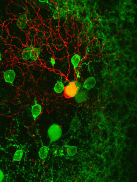
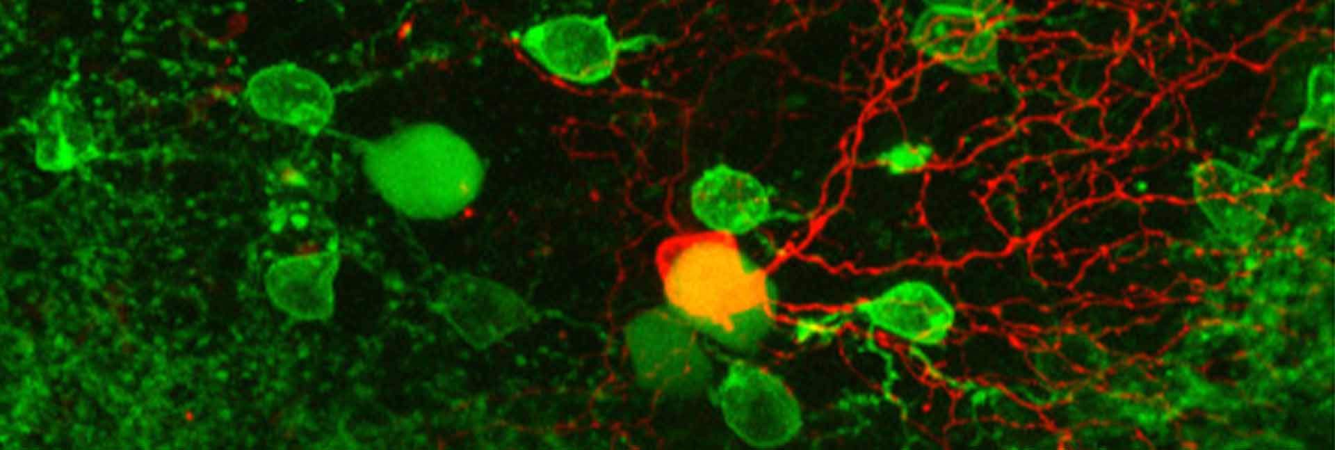
 and the starburst (green) cells surrounding that mediate the computation of motion direction")
Several years ago, when Dr. Michal Rivlin of the Institute’s Neurobiology Department was conducting postdoctoral research at the University of California, Berkeley, she discovered, to her surprise, that a certain type of light stimulus could get direction-selective retinal ganglion cells to reverse the direction of movement they responded to by 180 degrees. This was unexpected: The cells changed their direction selectivity despite the fact that the selectivity to motion is thought to be “hard-wired” via asymmetric connections from other neurons in their network. For example, direction-selective cells encoding left-to-right movement would receive inhibitory signals from cells to their right, and so on.
In the present study, led by research student Lea Ankri in Rivlin’s lab and conducted together with postdoctoral fellow Dr. Elishai Ezra-Tsur, the researchers set out to uncover what, exactly, happens to that neural circuit for sensing direction. Was the surprising reversal Rivlin had seen in her earlier research evidence of an unrecognized mechanism for perceiving movement?
The scientists used a combination of methods, including two-photon microscopy and electro-physiological recording in mice retina cells. This study revealed that the flip in direction was mediated by a change in the center-surround organization of the cells. “We found that certain visual stimuli masked the center of the cells’ visual field, causing the surround to take control of computing the signals,” says Ankri. “Surprisingly, the surround encodes for motion opposite to that encoded by the center of the field. We knew that antagonistic center-surround was an organizing principle that enabled better resolution for shape and color; to those we can now add motion.”
 Dr. Michal Rivlin and Lea Ankri")
Eyes are said to be the “windows of the soul,” but in Rivlin’s lab, the eyes’ retinas are windows into the brain. The human brain has some 100 billion nerve cells which have, between them, around 100 trillion electrical connections. This makes it difficult to draw even the most general conclusions about the “operating system” or its basic organizing principles. In contrast, the retina of a mouse eye has just a few million nerve cells, and, despite its complexity, many of its connections and circuits have been mapped out. Rivlin and her lab team, through their detailed approach to uncovering the computational processes in the retina, may be helping to understand similar processes within the brain.
Finding antagonistic center-surround motifs in these particular direction-selective cells suggests that the approach is a promising one. Antagonistic center-surround has been identified in higher-processing centers in the brain in addition to other sensory systems, and the researchers think it is probably a basic principle in neural computation that cuts across levels of complexity and function. It is the dynamic balance between center and surround responses that underlies the change in the directional responses of these cells, and this finding implies that the antagonistic organization is flexible. Such flexibility may be one of the things that enables our eyes – and our brains in general – to be so dynamic.
Dr. Michal Rivlin's research is supported by the Charles and David Wolfson Charitable Trust; the Rolf Wiklund and Alice Wiklund Parkinson’s Disease Research Fund; the Consolidated Anti-Aging Foundation; Dr. and Mrs. Alan Leshner; and Dr. Daniel C. Andreae. Dr. Rivlin is the incumbent of the Sara Lee Schupf Family Chair.