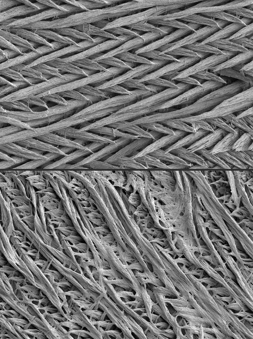
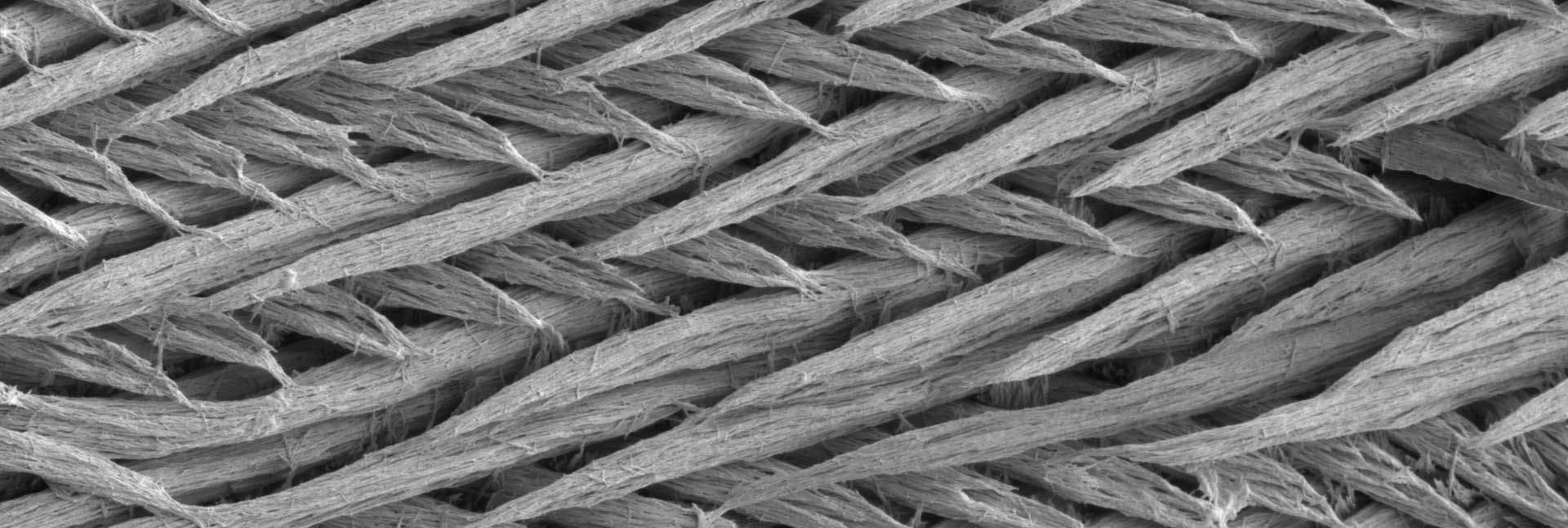
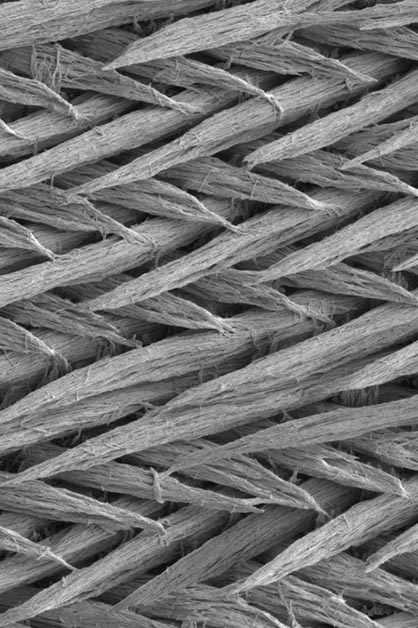
Enamel, the hardest and most mineral-rich substance in the human body, covers and protects our teeth. But in one of every 10 people – and in one third of children with celiac disease – this layer appears defective, failing to protect the teeth properly. As a result, teeth become more sensitive to heat, cold and sour food, and they may decay faster. In most cases, the cause of the faulty enamel production is unknown.
Now, a study by Prof. Jakub Abramson and his team at the Weizmann Institute of Science, published recently in Nature, may shed light on this problem by revealing a new children’s autoimmune disorder that hinders proper tooth enamel development. The disorder is common in people with a rare genetic syndrome and in children with celiac disease. These findings could help develop strategies for early detection and prevention of the disorder.

Tooth enamel is made up primarily of mineral crystals that are gradually deposited on protein scaffolds during enamel development. Once the crystals are in place, the protein scaffold is dismantled, leaving behind a thin but exceptionally hard layer that covers and protects our teeth. A strange phenomenon was identified in people with a rare genetic disorder known as APS-1: Although the enamel layer of their milk teeth forms perfectly normally, something causes its faulty development in their permanent teeth. Since people with APS-1 suffer from a variety of autoimmune diseases, Abramson and his team hypothesized that the observed enamel defects may also be of an autoimmune nature – in other words, that their immune system could be attacking their own proteins or cells that are necessary for enamel formation.
In general, autoimmune diseases occur when the immune system’s T cells or its antibodies mistakenly trigger an immune response against the body’s own cells or tissues. To prevent these incidents of “friendly fire,” T cells developing in the thymus gland need to first be educated to discriminate between the body’s own proteins and those of foreign origin. To this end, T cells are presented with short segments of self-proteins that make up various tissues and organs in the body. When a “poorly educated” T cell erroneously identifies a self-protein in the thymus as a target for attack, that T cell is labeled as dangerous and destroyed, so that it could not cause any damage after being released from the thymus.
""K-casein increases the amount of cheese that can be produced from milk, so the dairy industry deliberately raises its concentration. Our study, however, found that it may potentially trigger an immune response that can harm the body"
This critical education step is impaired in APS-1 patients as a result of a mutation in a gene known as the autoimmune regulator (Aire). This gene is essential for the T cell education process: It produces a protein that is responsible for the collection of self-proteins presented to the T cells in the thymus. In their new study, scientists from Abramson’s lab in Weizmann’s Immunology and Regenerative Biology Department, led by research student Yael Gruper, sought to work out how mutations in the Aire gene lead to deficient tooth enamel production. The researchers discovered that, in the absence of Aire, proteins that play a key role in the development of enamel are not presented to the T cells in the thymus gland. As a result, T cells that are liable to identify these proteins as targets are released from the thymus, and they encourage the production of antibodies to the enamel proteins. But why do these autoantibodies damage permanent teeth and not baby teeth?
The answer to this question lies in the fact that milk teeth develop in the embryonic stage, when the immune system is not yet fully formed and cannot create autoantibodies. In contrast, the development of enamel on permanent teeth starts at birth and continues until around the age of six, when the immune system is sufficiently mature to thwart enamel development. Furthermore, the researchers found a correlation between high levels of antibodies to enamel proteins and the severity of the harm to enamel development in children with APS-1. This strengthens the assumption that the presence of enamel-specific autoantibodies in childhood can potentially lead to dental problems.
When the researchers looked into deficiencies in enamel development in people with other autoimmune diseases, they found a very similar phenomenon in children with celiac disease, a relatively common autoimmune disorder that affects around 1 percent of people in the West. When people with this disease are exposed to gluten, their immune system attacks and destroys the cellular layer lining the small intestine, leading to attacks on other self-proteins in the intestine.

In an attempt to understand how celiac disease, known to cause intestinal damage, may also cause damage to tooth enamel, the researchers first examined whether people with this disease have autoantibodies that attack enamel. They found that a large proportion of celiac patients have these autoantibodies, just as do people with APS-1. But the “education” that takes place in the thymus gland of these patients seems normal, so why do they develop these antibodies? The researchers hypothesized that some proteins are found in both the intestine and the dental tissue and that these proteins play an important role in the development of tooth enamel. In this case, the antibodies that identify proteins in the intestine might move through the bloodstream to the dental tissue, where they could start to disrupt the enamel production process.
Since many celiac patients had previously been found to develop sensitivity to cow’s milk, the researchers decided to focus on the k-casein protein, a major component of dairy products. Strikingly, they found that the human equivalent of k-casein is one of the main components of the scaffold necessary for enamel formation. This led them to hypothesize that antibodies produced in the intestines of celiac patients in response to certain food antigens, such k-casein, may subsequently cause collateral damage to the development of enamel in the teeth, similarly to the way in which antibodies against gluten can eventually trigger autoimmunity against the intestine.
Indeed, they discovered that most of the children diagnosed with celiac had high levels of antibodies against k-casein from cows’ milk, which in many cases can also react against k-casein’s human equivalent expressed in the enamel matrix. This means that in theory, the same antibodies that are produced in the intestine against the milk protein could act against the human k-casein in the teeth.
These findings could have implications for the food industry. “Similarly to the lessons learned from gluten, we can assume that the consumption of large quantities of dairy products could lead to the production of antibodies against k-casein,” Abramson explains. “This protein increases the amount of cheese that can be produced from milk, so the dairy industry deliberately raises its concentration in cow's milk. Our study, however, found that the milk k-casein is a potent immunogen, which may potentially trigger an immune response that can harm the body itself.”
Tooth enamel flaws are common, not just among people with celiac disease or APS-1. “Many people suffer from impaired tooth enamel development for unknown reasons,” Abramson says. “It is possible that the new disorder we discovered, along with the possibility of diagnosing it in a blood or saliva test, will give their condition a name. Most important, early diagnosis in children may enable preventive treatment in the future.”


