Are you a journalist? Please sign up here for our press releases
Subscribe to our monthly newsletter:
Like sci-fi creatures in a B movie, many types of cancer cells grow protrusions from their bodies. Some are simply "feelers” for interacting with the cell’s surrounding. But one particular class of such protrusions – the aptly named invadopodia – can degrade the surrounding matter, disconnecting the cancer cells from the encasing tissues. These cells can then move to remote places in the body, leading to metastasis – the main cause of death from cancer. Prof. Benjamin Geiger and research student Or-Yam Revach of the Weizmann Institute’s Molecular Cell Biology Department have now revealed a new mechanism whereby invadopodia operate to promote the escape of cancer cells from their surroundings.
Geiger and his team knew that the invadopodia appear on cells in the course of interaction with the extracellular matrix – a dense, fibrous, adhesive medium that holds our body’s tissue together. To invade the matrix, invadopodia work in three steps. The cells first cling to the matrix around them through a so-called “adhesion domain.” They then degrade the matrix with special enzymes they produce. Finally, they push into the matrix with the help of structural materials from inside the cell. This common model for cancer cell invasion raises a question: How does the protrusion generate the force needed for penetrating the matrix? Geiger and Revach discovered a new mechanical model, which was recently published in Scientific Reports, that sheds new light, in particular, on this critical stage of this process.

The team first looked at the dynamic properties of the invadopodia adhesion sites, revealing that the adhesion domains are transient structures. Geiger compares this to an explorer trying to move inside of a thick bush, temporarily grabbing at vines and branches before cutting them and pushing his way in.
But the adhesion domains do not just disappear: A structure is assembled in their place out of microtubules made of the cytoskeleton support proteins. This assembly supports the developing invadopodia and stabilizes them.
In the new model, the invadopodia function something like a drill.The concept of a drilling mechanism that pushes mechanically raised a new question: How does this drill-like structure brace itself? An explorer in the bush and a person drilling into a wall are both braced by the same thing: the ground. The hardness of the ground beneath our feet gives us the counterforce we need to propel ourselves forward or apply force to another material. But the cell has no such “hard ground” to provide that counterforce. And the extracellular matrix has about the same squishy consistency as the cell itself, so it cannot provide the mechanical support needed for drilling or moving forward.

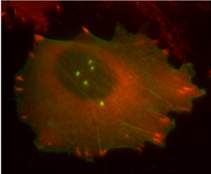
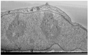
A closer look at the microtubule edifice showed that the structure inside the protrusion is just the tip: It extends into the cell – all the way to the nucleus, where it dents the nuclear walls. In other words, it looked as though the cell’s nucleus, a walled compartment that safeguards the cell’s genetic material, could be providing the counterforce needed for drilling into the matrix. To obtain this closer look, the scientists developed a technique that enabled them to visualize invadopodia, first by light microscopy, then “zooming-in” to determine the three-dimensional structure by high-resolution electron microscopy and, finally, precisely matching up the images from both types of microscopy.
Another member of the group, Dr. Ariel Livne, then calculated the force applied by the invadopodia cytoskeletal core on the indented nucleus, on the one side, and on the matrix, on the other. The group then looked at the molecules that are involved in the process: When they interfered with the assembly of the invadopodia structures, the result was flat nuclear walls; blocking the proteins that link the nucleus to the cytoskeleton resulted in long-lasting adhesion domains, suggesting that the adhesion domains compensate for the lack of interaction at the invadopodia’s nuclear tip.
