Within minutes of fertilization, the egg of a fruit fly becomes a scene from the battle of the sexes. The egg attacks and destroys the cellular “power plants,” or mitochondria, from the sperm that had fertilized it, so that only its own mitochondria remain. These findings from a new Weizmann Institute of Science study, published in Nature Communications, might provide insights into advanced fertilization treatments and shed light on a long-standing mystery: How and why do we inherit all of our mitochondria from our mothers?
Paternal mitochondria vanish soon after fertilization in virtually all species, from sexually reproducing plants through fungi and insects to mammals, including humans. One theory for this holds that paternal mitochondria are simply diluted by the vastly more abundant maternal ones, but another suggests that they are actively eliminated by the egg.
About a decade ago, Prof. Eli Arama’s lab in Weizmann’s Molecular Genetics Department provided crucial evidence in favor of the active elimination theory. The new study, led by PhD student Sharon Ben-Hur, reveals the molecular details of this elimination, showing that the egg launches a deliberate, targeted attack on the sperm’s mitochondria.
“It’s possible that paternal mitochondria carry harmful components, but they might even contain certain gifts from the father,” Arama says. “Either way, breaking them down may crucially affect embryonic development.”
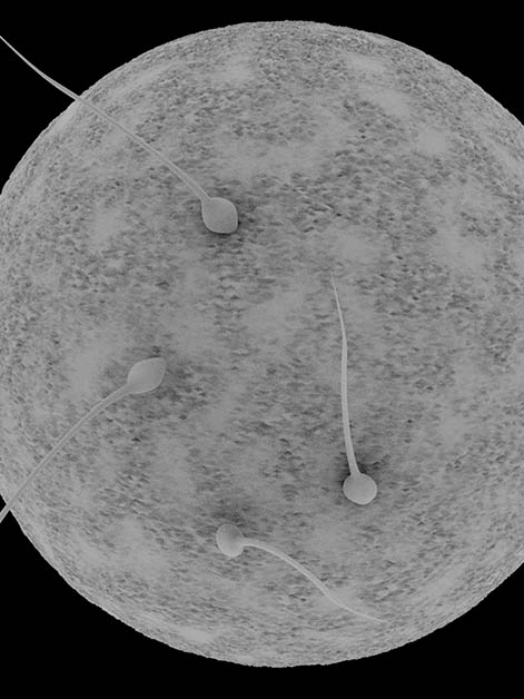
The sperm of the fruit fly, or Drosophila, is one of the largest in nature, making it an excellent model for these studies. Its mitochondria are not scattered, but rather fused along its tail into a single elongated structure. Yet the fruit-fly egg, as the team showed, manages to destroy even this large formation.
Ben-Hur and colleagues found that no sooner does the sperm penetrate the egg than it is greeted by swarms of vesicle clusters. In a manner that is obviously purposeful and preprogrammed, these clusters immediately fuse to form a vesicular sheath covering the entire length of the sperm’s tail. Next, the mitochondrial structure in the sperm’s tail is broken down, sheath and all, into smaller chunks.
If the findings are confirmed in further studies, they might help improve state-of-the-art fertility treatments
After studying thousands of fruit-fly embryos, Ben-Hur and team were surprised to discover that the vesicles forming the sheath contain molecules that are involved in innate immunity. Further research showed that the outer surface of the vesicular sheaths harbors large amounts of a protein called Rubicon, which is known to play a role in an immune pathway called LC3-associated phagocytosis or, for short, LAP, known to function against invading microbes.
This insight led the researchers to decipher the entire pathway involved in the mitochondria’s degradation and to reveal that it is indeed similar to LAP. Furthermore, just as happens inside immune cells that target microbes, LAP’s final step in the egg involves the recruitment of lysosomes, the cell’s recycling organelles, which complete the degradation of the mitochondria.
“We discovered that the egg repurposes an innate immunity pathway to destroy paternal mitochondria. In a way, it treats them as dangerous trespassers,” Ben-Hur says. This discovery of the egg’s recourse to antimicrobial mechanisms fits with the prevailing hypothesis regarding the mitochondria’s primordial origins: In the ancient past, the mitochondria might have started out as bacteria that invaded a higher organism’s cells and stayed on because the invasion proved beneficial to both sides.
By explaining how the egg handles the destruction of such enormous constructs as the fruit fly’s paternal mitochondria, the study might open a new research direction in cellular biology. Its findings could guide a search for previously unknown ways in which cells degrade large structures such as entire damaged organelles.
The study might also offer new clues as to why paternal mitochondria need to be destroyed. One common explanation relates to the cell’s need for maintaining compatibility between its two genomes: one in the nucleus, which results from a merger of maternal and paternal DNA, and another, different one in the mitochondria. According to this explanation, such compatibility should be easier to achieve when all the mitochondria carry only maternal DNA, since too many different DNAs might clash. That might not be the whole story, however. Paternal mitochondria are vastly outnumbered by maternal ones, and their breakdown in both fruit flies and humans occurs long after their DNA has been eliminated.
“The fact that the egg resorts to surprisingly aggressive mechanisms to destroy paternal mitochondria suggests an urgency,” Arama says. “One possible reason is that these mitochondria might contain certain non-DNA components, such as RNAs, that are deleterious to the embryo – or, on the other hand, small non-genetic molecules that are made available by the mitochondria’s destruction and might be vital to the embryo’s development.” In fact, when Ben-Hur created mutant fly embryos without the Rubicon protein, these embryos failed to destroy paternal mitochondria – and failed to develop properly.
Are paternal mitochondria in humans and other mammals destroyed by the same mechanisms as in the fruit flies? Certain similarities have already emerged, including the expression of the LC3 molecule on paternal mitochondria and the presence of vesicle clusters in the vicinity of the mammalian sperm tail after fertilization.
If these similarities are confirmed in further studies, they might help improve state-of-the-art fertility treatments. For example, in one common IVF technique, a single sperm is injected into the egg to increase the likelihood of fertilization, instead of exposing the egg to multiple sperm in a test tube. However, evidence from other areas of cellular biology suggests that an injected sperm might lack the markers needed for the destruction of its mitochondria – markers it would have acquired had it penetrated the egg spontaneously. Adding these markers could potentially contribute to the treatment’s success.
Other fertility treatments involve replacing the egg’s mitochondria with those from a donor, for example, when maternal mitochondria are known to carry a disease-causing mutation. In such cases, understanding the mechanisms of mitochondrial destruction can help ensure that the donor mitochondria are properly integrated into the fertilized egg.
Science Numbers
The tail of a fruit fly sperm is about 1.8 millimeters long – approximately 30 times longer than that of the human sperm, which measures about 60 microns (micrometers) in length.
A mature mammalian egg has between 100,000 and 600,000 mitochondria, compared to only 50 to 75 mitochondria in a single mammalian sperm.
Study participants also included Shoshana Sernik, Sara Afar, Dr. Alina Kolpakova, Dr. Yoav Politi, Dr. Liron Gal, Dr. Anat Florentin, Prof. Shmuel Pietrokovski, Dr. Eyal Schejter and Dr. Keren Yacobi-Sharon of the Molecular Genetics Department; Ofra Golani and Dr. Ehud Sivan of the Life Sciences Core Facilities Department; Dr. Nili Dezorella of Chemical Research Support Department; and Dr. David Morgenstern of the Nancy and Stephen Grand Israel National Center for Personalized Medicine.
Prof. Eli Arama is head of the Crown Human Genome Center and the incumbent of the Harry Kay Professorial Chair of Cancer Research. His research is supported by the Kekst Family Institute for Medical Genetics.


 Dr. Keren Yacobi-Sharon, Shoshana Sernik, Prof. Eli Arama, Sharon Ben-Hur, Dr. Eyal Schejter and Dr. Alina Kolpakova")
 are clearly visible in the egg several minutes after it’s been laid (top left), but are completely destroyed within an hour (top, second from left). In a mutant fly lacking the Rubicon protein (bottom left), these mitochondria are not destroyed, even 1, 2 or 3 hours after egg laying (bottom row, second, third and fourth from left)")
 encased by a sheath containing abundant Rubicon protein (green)")