A few well-known facts about lasers: These ultra-focused beams of light have been used for decades to cut metal cleanly and precisely. In the field of medicine they take the place of sharp surgical scalpels; they are also used in some kinds of medical imaging; and they are a part of today’s advanced optical microscopes. Many of these uses require the light to be focused tightly to a very narrow, highly intense point. One way of cutting cleanly without causing harm to the surrounding area – say, in biological tissue that is easily damaged by excess energy – is to zap the target point with very brief (less than a millionth of a millionth of a second) flashes of highly concentrated laser light.
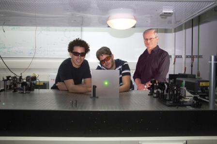
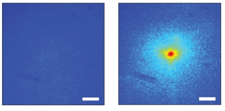
Prof. Yaron Silberberg and research students Ori Katz, Eran Small and Yaron Bromberg of the Physics of Complex Systems Department sought a way to focus rapid flashes of laser light as they pass through a scattering layer. The method they developed works on feedback: They created a system that can assess, in real time, how the light scatters. Using algorithms they developed, they created a beam that can “anticipate” the dispersal of its light and make the necessary corrections. This computerized system is able to tailor a laser beam to the tissue such that it is precisely and narrowly focused on the internal target.
In this research, which appeared in
Nature Photonics, the scientists employed a simple LCD screen, similar to those found in computer projectors, to correct the beams’ focus. While it was known that screens of this type could be used to correct spatial errors, Silberberg and his team succeeded in demonstrating that this simple system can also correct for errors in the dimension of time.
The scientists hope that this system will, in the future, aid in the development of applications, including new types of medical lasers and optical microscopes that will enable researchers in the life sciences to get under the skin and into the underlying tissue.
Prof. Yaron Silberberg’s research is supported by the Crown Photonics Center, which he heads; the Wolfson Family Charitable Trust; and the Cymerman - Jakubskind Prize. Prof. Silberberg is the incumbent of the Harry Weinrebe Professorial Chair of Laser Physics.

