Are you a journalist? Please sign up here for our press releases
Subscribe to our monthly newsletter:
Whenever we lay eyes on a mouth-watering dessert, swallow a piece of food or start crying, cells in our bodies release specialized fluids: saliva, digestive juices or tears. Ways in which these fluids and other substances move to the outsides of the cells have been studied for nearly a century – they are the stuff of biology textbooks. But the Weizmann Institute of Science’s Dr. Ori Avinoam and Prof. Ben-Zion Shilo realized that something was not quite right about the textbook descriptions.
Exocytosis, the release of substances from cells, occurs through tiny bubbles called vesicles that fuse with the cell’s surface, becoming part of the cellular membrane and spilling their contents to the outside. In this manner cells expel waste products and dispatch various signaling molecules, including hormones and neurotransmitters. For example, when neurons communicate with one another by means of neurotransmitters, neurotransmitter-containing vesicles fuse with the neuronal membranes, spilling the substances outside for adjacent neurons to sense. This wave of exocytosis is followed by a mirror-image process, in which the vesicles are “recycled”: The neurons take the neurotransmitter back in, inside new vesicles created from the ones that had fused with their membranes. As a result, the vesicles and the cellular membranes go back to their original state, enabling the neurons to function over and over.
This classic picture accurately depicts the release of substances from vesicles measuring less than 100 nanometers (billionths of a meter) across, in a burst that lasts just milliseconds. But cells known as “professional secretors,” whose job it is to secrete, or produce digestive enzymes, moisture and lubricants in various glands and tissues – such as the salivary and tear glands, and the lining of the mouth or lungs – package their cargo in vesicles that are as much as a hundred times larger in diameter, measuring up to 10 microns (millionths of a meter) across. These vesicles discharge their contents over a period of several minutes. In terms of the ratio of surface area to volume, they are much more economical than small vesicles for releasing large amounts of material. Still, their surface is so extensive that once they fuse with the cellular membrane, the cell could swell and become completely distorted.
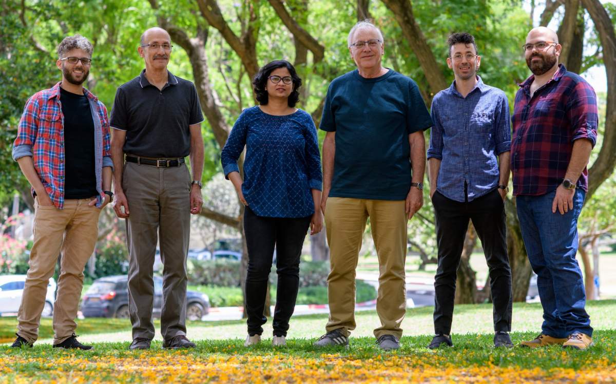
Shilo, of the Molecular Genetics Department, and Avinoam, of the Biomolecular Sciences Department, discussed this conundrum. Shilo’s lab had been studying salivary glands in fruit fly larvae as a model for secretion by giant vesicles. Avinoam’s new lab explores the mechanisms of membrane remodeling, with a focus on advanced imaging techniques. The two came to the conclusion that a central unresolved issue in the secretion process from giant vesicles is how the cells manage to keep their membranes intact. Fusion of multiple giant vesicles with this membrane along the lines proposed by the classic model would inevitably make the cell completely dysfunctional.
To resolve the mystery, Shilo and Avinoam teamed up with Dr. Kamalesh Kumari, a joint postdoctoral fellow in the two labs, who led the study. The scientists used 3D electron microscopy and other imaging methods to examine cells in the salivary gland of fruit fly larvae and in the mouse exocrine pancreas, both of which secrete substances through giant vesicles. The research team included Nadav Scher, Tom Biton and Dr. Eyal D. Schejter.
“Our study revealed that giant vesicles, in contrast to the small ones, release substances from cells via a previously unknown mechanism – one that’s completely different from the textbook scenarios,” Avinoam says.
"Defects in the crumpling mechanism might very well be involved in diseases in which the release of fluids from cells is insufficient, for example, dry eye disease, or alternatively, is excessive, as happens in cystic fibrosis"
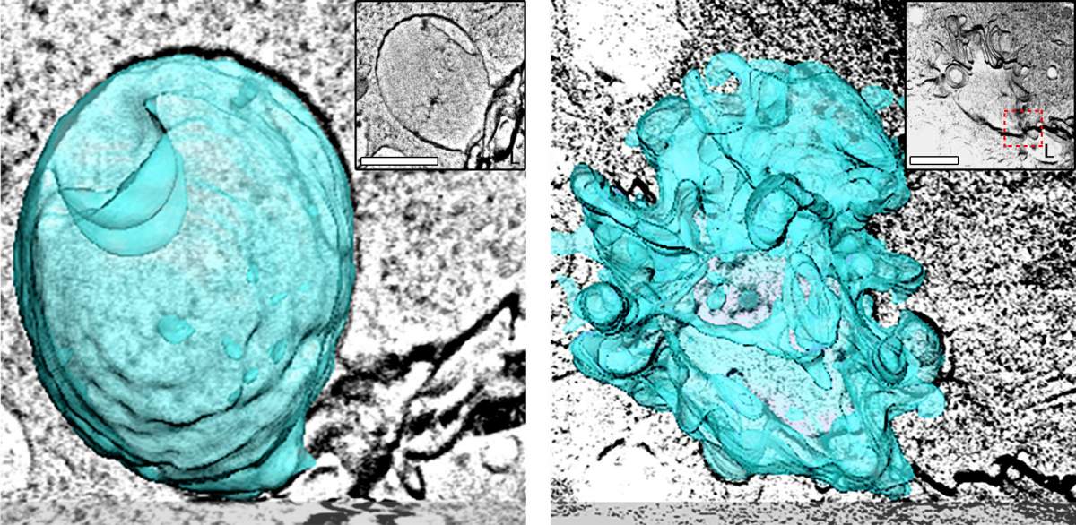
It turned out that when a large vesicle fuses with the cellular membrane, it does not become integrated into the cell’s surface. Rather, it is enveloped by a protein network called actomyosin, which works like a toothpaste squeezer or a turkey baster, gradually squeezing the vesicle’s contents outside the cell through a small opening. In the course of the squeeze, the actomyosin crumples the vesicle’s wall – much the way you might crumple a piece of paper to make a toy ball for a cat – while at the same time keeping the vesicle physically distinct from the cell membrane.
This setup ensures that the cellular membrane is not disrupted in the process. As for the empty crumpled vesicle, it recruits molecular machinery that recycles its wall, producing thousands of small vesicles that serve to move substances to the inside of the cell.
“These findings open up entirely new directions for studying the release of material from giant vesicles, particularly the mechanisms that maintain the integrity of cellular membranes during exocytosis,” Shilo says.
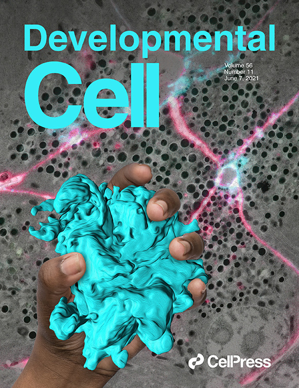
The fact that exocytosis by crumpling was observed not only in fruit fly larvae but also in mouse cells, which more closely resemble human secretory tissue, suggests that this mechanism is likely to be at work in human cells as well. And this, in turn, points to new angles for exploring potential causes of various human diseases.
“Defects in the crumpling mechanism might very well be involved in diseases in which the release of fluids from cells is insufficient, for example, dry eye disease, or alternatively, is excessive, as happens in cystic fibrosis,” Kamalesh says. “Such defects may also play a role in disorders affecting the body’s inner surfaces that need to be lubricated properly in order to function well, for example, the inner lining of the digestive tract.”
Ori Avinoam made the switch from student to teacher for the first time while still in high school in Haifa. When his biology teacher fell ill during preparations for the matriculation exams, other students asked Avinoam to take over that day, much to the teacher’s delight. “Biology always came naturally to me,” he recalls.
While studying for a BSc in Biology and Biochemistry at the Technion – Israel Institute of Technology, Avinoam became fascinated by biological membranes, which are essential for the life of every cell. He started looking for a summer internship that would help him understand the interplay between membranes and the proteins that populate them.
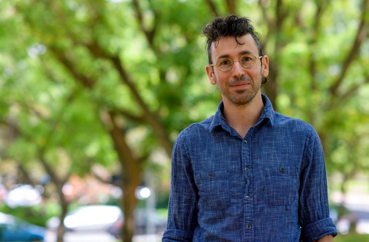
In the lab that he eventually joined, he was struck by glowing cells viewed with fluorescence imaging, then a relatively new approach in biology. “I saw luminous circles that moved,” Avinoam says. “They turned out to be the nuclei of cells in the body of a transparent microscopic worm. This totally blew my mind.”
In his studies on a direct PhD track at the Technion, and later in postdoctoral studies at the European Molecular Biology Laboratory in Heidelberg, Germany, Avinoam found a way to combine the magic of both fields. He embarked on a study of membranes using advanced imaging methods.
As a young scientist, Avinoam recalls being particularily struck by images of glowing cells viewed with fluorescence imaging, then a relatively new approach in biology: "This totally blew my mind"
During his postdoctoral studies, he made yet another student-to-teacher switch. At the request of his colleagues, he started teaching them yoga, which he had been practicing for a number of years. “I find a great deal of similarity between practicing yoga and doing science – both are rooted in reflection,” Avinoam says.
In his lab at Weizmann, Avinoam studies cellular membranes using fluorescence and electron microscopy, as well as other state-of-the-art methods. He focuses on processes involved in cell-to-cell fusion and in the movement of various types of molecular cargo from one membrane compartment to another, such as those enabling the fusion of sperm and egg, the development of skeletal muscles or the release of materials from cells through vesicles.
And once a week, he teaches a yoga class to his lab members and friends.
Avinoam lives on campus with his partner Yuval, an aeronautics engineer, and their three-year-old twins, a boy and a girl.

The combined surface area of all large vesicles within a pancreatic cell that secretes digestive enzymes is 30 times greater than the area of the cell's membrane to which they become attached.
Dr. Ori Avinoam is the incumbent of the Miriam Berman Presidential Development Chair.
Dr. Avinoam's research is supported by the the Yeda-Sela Center for Basic Research; the Henry Chanoch Krenter Institute for Biomedical Imaging and Genomics; and the Schwartz/Reisman Collaborative Science Program.
Prof. Ben-Zion Shilo is the incumbent of the Hilda and Cecil Lewis Professorial Chair of Molecular Genetics.
Prof. Shilo's research is supported by the Henry Chanoch Krenter Institute for Biomedical Imaging and Genomics; the Yeda-Sela Center for Basic Research; and the Braginsky Center for the Interface between Science and the Humanities.