Are you a journalist? Please sign up here for our press releases
Subscribe to our monthly newsletter:

Saliva, teardrops, bile – all kinds of vital bodily fluids are produced in specialized cells whose sole function is to secrete one particular substance on demand. These cells are essentially closed, walled systems. How are they able to move large amounts of these substances to the outside? Surprisingly, researchers have not had a good answer to this question until now. Dr. Tal Rousso, a postdoctoral researcher in the group of Prof. Ben-Zion Shilo in the Molecular Genetics Department, looked closely at the process in a unique model – fruit fly larva salivary glands, which secrete a sort of glue – and discovered that these cells have an interesting way of squeezing out the fluids they produce. The group’s findings appeared in Nature Cell Biology.
For their size, explains Shilo, fruit fly larvae have very large salivary glands, and the secretory cells lining these glands are exceptionally large. Those cells deposit a sort of glue into the gland, and when a larva is ready to undergo metamorphosis into an adult fly, it “spits” the glue onto a leaf and sticks itself in place as it becomes a pupa.
Secretion takes place with the help of special bubble-like compartments called vesicles that envelop the substance in a membrane and transport it to the outer membrane of the cell. Here the vesicle fuses with the membrane, opening up to the outside. Many cellular substances are expelled through vesicles, and this process has been well studied, but “professional” secretory vesicles, which are thousands of times larger than the ordinary vesicles in an average cell, raised a new set of questions. For example, how does the vesicle know how to get to the part of the membrane that faces the lumen – the collection vessel? And how does it efficiently expel its bulky package – especially a viscous one like saliva or glue?
When a larva is ready to undergo metamorphosis into an adult fly, it “spits” the glue onto a leaf and sticks itself in place
Rousso, Shilo and Dr. Eyal Schejter were able to observe the living cells lining the lumen in the fruit fly salivary glands, using a type of time-lapse photography that they developed in their lab. They first noted that as more and more vesicles spill their contents into the lumen, filling it up, the cells shrink back. This effectively brings the cell membrane in toward the vesicles. Still, the researchers noted that the vesicles are physically attracted to the inside membrane – that facing the lumen – as this part is constructed differently from the sides and outside-facing walls of these cells. The scientists’ observations showed that at the membrane surface, each vesicle gets coated with two materials – actin and myosin – that help to actively squeeze the substance out into the lumen. The actin forms tiny tracks, and the myosin molecules are motors that attach to the tracks and cause the vesicle to contract. The myosin motors pull on the actin tracks in various random directions, but the overall their pulling is to bring the tracks closer together.
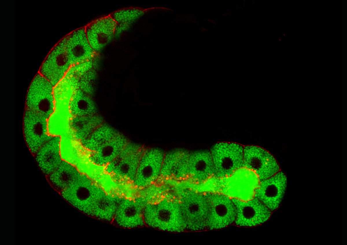
What causes this coating to form? To answer this question, Rousso infused red dye into the lumen and labeled the actin with green fluorescent proteins. The time-lapse films showed that the red beads infused into the vesicles just before the green coating formed, proving that the vesicle first fuses with the membrane and begins to open; it is this fusion that initiates the next stage. They then identified a protein that is transferred to the vesicle membrane and attracts both the actin and the myosin.
Further observation revealed that the myosin works in a surprising way: It concentrates in stripes like those on a basketball. It turns out that these stripes are self-organizing; the stripes then contract like fingers squeezing the vesicle. A comparison with vesicles whose contraction machinery was distributed equally over the surface revealed that the former is the more efficient way to expel the vesicle contents.
“Our bodies have many different kinds of large secretory cells,” says Shilo. “This research was an opportunity to understand how they work, live and up close.”
Glue protein secretion from larval salivary glands. Time-lapse movie of cultured fruit-fly salivary glands. Actin coating of the vesicles shows up in fluorescent red, and the glue is seen in fluorescent green. Over time, the lumen (center) fills with glue and the cells, which have expelled their contents, shrink
Prof. Ben- Zion Shilo's research is supported by the M.D. Moross Institute for Cancer Research; and the Mary Ralph Designated Philanthropic Fund. Prof. Shilo is the incumbent of the Hilda and Cecil Lewis Professorial Chair of Molecular Genetics.