WOWing the Crowds

English
 
A team of scientists at the Weizmann Institute of Science and the Hebrew University of Jerusalem has developed a method that could speed up the process of identifying novel protein molecules hundreds of times over.

Instead of plates containing rows of tiny wells, the new method - developed by Drs. Dan Tawfik and Amir Aharoni of the Institute's Biological Chemistry Department and Prof. Shlomo Magdassi of the Hebrew University's Institute of Chemistry, with support from the Israel Ministry of Science and Technology - relies on microscopic droplets of water suspended inside oil droplets. The method, which uses an emulsion dubbed WOW (water-oil-water), takes a lead from living cells, which employ a fatty membrane to keep their inside and outside environments separate. Using the new system, millions of tests can be performed at once.

The method involves adding a fluorescent marker that lights up in color when activated by the right protein and sorting through the droplets for those containing the marked proteins and their coding genes. Automated devices for sorting cells can handle many thousands of droplets per second. "Searches that now take a year to complete could be done in a matter of days," says Tawfik.  

 
Dr. Dan Tawfik's research is supported by the Y. Leon Benoziyo Institute for Molecular Medicine; the Dolfi and Lola Ebner Center for Biomedical Research; the Estelle Funk Foundation; the Dr. Ernst Nathan Fund for Biomedical Research; the Henry S. and Anne Reich Family Foundation; the Charles and M.R. Shapiro Foundation Endowed Biomedical Research Fund; the Harry and Jeanette Weinberg Fund for the Molecular Genetics of Cancer; the Eugene and Delores Zemsky Charitable Foundation; and Mr. and Mrs. Mordechai Segal, Israel. Dr. Tawfik is the incumbent of the Elaine Blond Career Development Chair.
Technology & Applications
English

Living with Salt

English
 
Life thrives in all sorts of hostile environments, including the extreme salinity of the Dead Sea. For over 30 years, Weizmann Institute scientists have been investigating how Dunaliella salina, a microscopic, plant-like alga, is able to proliferate in such inhospitable surroundings. As an offshoot of these and other studies, Dunaliella is today commercially grown as a source of natural beta carotene. Now Institute scientists have unraveled one of the secrets of the alga’s exceptionally successful adaptation to salt that, unexpectedly, might also shed light on the working of our own kidneys.
 
Prof. Ada Zamir and Dr. Lakshmanane Premkumar of the Biological Chemistry Department and Prof. Joel Sussman and Dr. Harry Greenblatt of the Structural Biology Department focused on an exceptionally salt-tolerant Dunaliella enzyme, a carbonic anhydrase. They showed that although the Dunaliella enzyme shares a basic plan with salt-intolerant carbon anhydrases from animal sources, it has unique structural features that enable it to remain functional in any degree of salinity, from the extremes of the Dead Sea to nearly fresh water.
 
In a surprising twist, the researchers discovered that one other carbonic anhydrase – found in mouse kidneys – sported a similar, salt-tolerant structure. Since the mouse enzyme closely resembles that of humans, the researchers hope that their findings might provide the basis for designing new drugs that could target carbonic anhydrase enzymes on the basis of their salt tolerance.
 

Human (left), Dunaliella (center) and mouse (right) enzymes


Prof. Joel Sussman’s research is supported by the Helen and Milton A. Kimmelman Center for Biomolecular Structure and Assembly; the Joseph and Ceil Mazer Center for Structural Biology; the Divadol Foundation; the Jean and Jula Goldwurm Memorial Foundation; Sally Schnitzer; the Kalman and Ida Wolens Foundation; and the Wolfson Family Charitable Trust. Prof. Sussman is the incumbent of the Morton and Gladys Pickman Professorial Chair in Structural Biology.
 
Human (left), Dunaliella (center) and mouse (right) enzymes
Chemistry
English

First Matter

English


The first matter that came into being right after the Big Bang may not have been quite what scientists had expected. This conclusion emerged recently from the large-scale PHENIX experiment carried out in the United States by a team of 460 physicists from 12 countries, including scientists from the Weizmann Institute.


Using an accelerator at the Brookhaven National Laboratory on Long Island, New York, the scientists may have recreated what’s believed to have been the primordial matter in the universe, known as the quark-gluon plasma. Two beams of gold ions were accelerated toward each other, causing head-on collisions. The enormous power of the collisions – about 40 trillion electron volts – turned part of the beams’ kinetic energy into various particles (a process described by Einstein’s E=mc2 equation). A number of the particle detectors used in the experiment were designed and built by Prof. Itzhak Tserruya and his team in the Weizmann Institute’s Particle Physics Department.


While many of the experimental results fit in with predictions of how particles in the quark-gluon plasma should behave, others have been a surprise. In particular, scientists were amazed to discover that the plasma, created at a heat up to 150,000 times hotter than the center of the sun, behaves not like a super-hot gas, as expected, but more like a liquid! The experiment, now in its fifth year, is scheduled to run for at least five more years and is certain to yield more susprises in the future.


Prof. Itzhak Tserruya’s research is supported by the Nella and Leon Benoziyo Center for High Energy Physics and the Center for Scientific Excellence. Prof. Tserruya is the incumbent of the Samuel Sebba Professorial Chair of Pure and Applied Physics.

Space & Physics
English

A Long and Winding Road

English


Long-distance messengers star in many heroic tales. A team of Weizmann Institute researchers has now discovered how molecular messengers help injured nerve cells to heal themselves.


Nerve cells in the peripheral nervous system can regrow when their extensions, called axons, are damaged. The injured axon issues a call for help, which is transmitted by molecular messengers that bind to molecules of phosphorus. In this phosphorylated state, they deliver a message that prompts the cell to manufacture proteins vital for the healing of the injured axon. The problem is that the messengers can easily lose their phosphorus message in the course of their arduous journey along the axon, which in the human body can reach up to a meter in length.


Dr. Michael Fainzilber and graduate students Eran Perlson and Shlomit Hanz, all of the Biological Chemistry Department, discovered that the molecular messengers, together with their phosphorus message, bind to a special molecule called vimentin, which in turn links up to motor proteins in the axon. It is thanks to this linkage and protection that the messengers can safely carry out the task of transmitting the axon’s call for help to the cell body. The scientists hope that these findings might advance the search for new therapies for injured nerve fibers.


The research team also included Prof. Rony Seger of the Biological Regulation Department, Prof. Michael Elbaum of the Materials and Interfaces Department, graduate students Keren Ben Yaakov and Yael Segal-Ruder of the Biological Chemistry Department and postdoctoral fellow Dr. Daphna Frenkiel-Krispin of the Materials and Interfaces Department.


Dr. Michael Fainzilber’s research is supported by the Y. Leon Benoziyo Institute for Molecular Medicine; the Nella and Leon Benoziyo Center for Neurological Diseases; the Irwin Green Alzheimer’s Research Fund; the Abisch Frenkel Foundation for the Promotion of Life Sciences; the Buddy Taub Foundation; and Mr. and Mrs. Alan Fischer. Dr. Fainzilber is the incumbent of the Daniel E. Koshland Sr. Career Development Chair.

Space & Physics
English

Ironing Genes

English

High levels of Ferritin show up in red, low levels in blue


An iron storage molecule in the cell, with the help of magnetic resonance imaging (MRI), can serve as an advanced tool for mapping gene expression.
 
Prof. Michal Neeman of the Weizmann Institute's Biological Regulation Department, together with Dr. Batya Cohen of the Molecular Genetics Department, altered the iron-bearing ferritin molecule to serve as a sort of gene “spy.” The scientists rendered ferritin sensitive to tetracycline, a common antibiotic, which serves as a switch, turning ferritin ON or OFF. As MRI is sensitive to magnetic particles such as iron, the cells' increased uptake of iron when ferritin is turned ON can be tracked, effectively exposing the genetically modified cells.
 
This method grew out of a joint vision that originated 10 years ago in a collaboration with the late Dr. Yoav Citri. It has far-reaching implications for monitoring the progress of gene therapy and for tracking gene expression in the central nervous system or during embryonic development.
 
Prof. Michal Neeman’s research is supported by the M.D. Moross Institute for Cancer Research; the Willner Family Center for Vascular Biology; the Mark Family Foundation; and Mr. and Mrs. Stephen Meadow.
 
 
MRI image: High levels of Ferritin show up in red, low levels in blue
Life Sciences
English

Transistor Genetics

English

Take a few tiny spoonfuls of phosphates, sugars and nucleotides to create several types of DNA, add a pinch of carbon nanotubes, sprinkle in a few grains of gold, mix well on a clean silicon surface -and what do you have? A transistor, according to research conducted by Prof. Ron Naaman of the Weizmann Institute's Chemical Physics Department.
 

Several unique strands of DNA were created and programmed to form different types of attachments. One tiny set of DNA strands was designed to connect to minuscule electrical contacts made of gold that were anchored to the silicon surface, while a second type of DNA strand was designed to fasten to the carbon nanotubes – extra-strong hollow tubes a mere 10 hydrogen atoms in diameter. The end result was a sort of carbon nanotube “bridge” spanning the silicon surface between two gold contacts.
 
Similar nanobridges may one day form the basis of tiny nanotransistors that will be used to build fast, efficient, miniaturized electronic circuits. This “recipe for success” appeared in Applied Physics Letters.  

 
Prof. Ron Naaman’s research is supported by the Fritz Haber Center for Physical Chemistry; the Ilse Katz Institute for Material Sciences and Magnetic Resonance Research; the Wolfson Advanced Research Center; the Philip M. Klutznick Fund for Research; and Dr. Pamela Scholl, Northbrook, IL. Prof. Naaman is the incumbent of the Aryeh and Mintze Katzman Professorial Chair.
 

DNA/nanotube bridge


A carbon nanotube (shown in brown) 
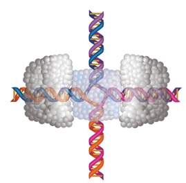
forms a bridge between two segments of DNA supported by gold contacts (yellow) attached to a silicon surface (green)
Chemistry
English

Nanotubes Stay in Step

English

Carbon nanotube on sapphire substrate


Sometimes a rough start makes for a smooth finish. Carbon nanotubes are excellent candidates for components of tiny nanoelectronic circuits, but their organization into ordered arrays on surfaces remains a major obstacle.


In an effort to manipulate their growth, the research group of Dr. Ernesto Joselevich of the Institute’s Materials and Interfaces Department experimented with the application of electrical fields to control the direction of nanotube formation on various surfaces. When a sapphire surface was chosen, the group was surprised to observe a beautiful parallel arrangement of nanotubes formed in an orientation completely independent of the electrical field. Closer examination of the sapphire revealed that it had not been cut precisely along the crystal plane, so the surface consisted of a series of terraces separated by steps of atomic dimensions. The iron nanoparticles used as a catalyst to produce nanotubes proved to be a lazy partner that didn't like to “climb stairs” but instead preferred to “glide” along the edge of the step. In its wake lay an organized trail of nanotubes formed snugly along this edge.  The nanotubes even followed kinks in the steps caused by defects in the crystal. This resulted in either straight or zigzag-shaped tubes, which are expected to have particularly interesting electronic properties.
 
“The direction and shape of the atomic steps can be controlled simply by the cut of the crystal,” says Joselevich. “This could lead to intriguing nanotube architectures.” Exploiting these small steps could mean a big step toward large-scale nano-fabrication to produce different nanowire arrangements in a controlled fashion.
 
Full details of the study appear in the cover story of the prestigious journal Angewandte Chemie
 
Dr. Ernesto Joselevich’s research is supported by the Ilse Katz Institute for Material Sciences and Magnetic Resonance Research; the Asher and Jeannette Alhadeff Research Award; Sir Harry A.S. Djanogly, CBE, UK; the Philip M. Klutznick Fund for Research; and Sylvia and Henry Legrain, Spain. Dr. Joselevich is the incumbent of the Dr. Victor L. Ehrlich Career Develop-ment Chair.
Nanotube formation along atomic steps
Chemistry
English

Cancer Therapy Penetrates Bone

English
 
When prostate cancer, one of the most lethal cancers, spreads in the body, it most often targets bone. Difficult to treat, such metastasis is implicated in over 70% of prostate cancer deaths. A new therapy crosses bone barriers.
 
Prof. Zelig Eshhar, Head of the Immunology Department at the Weizmann Institute of Science, previously developed prostate-cancer-fighting cells, dubbed T bodies, which are modified immune system cells customized to be highly effective in identifying and destroying cancer cells. However, T bodies were unable to effectively penetrate bone. The Weizmann team, which included Dr. Jehonathan Pinthus of Sheba Medical Center, Tel Hashomer, implemented a pre-treatment consisting of either low doses of radiation or a specific chemotherapy drug, followed by T body injections. The pre-treatments caused some disruption in the bone marrow, the intended target of the T bodies, which responded with a chemical distress signal that alerted the immune cells, aided them in locating the problem area and enabled them to pass through barriers into the bone marrow tissue.
 
Mice treated with either therapy showed a significant drop in the tumor marker PSA (an indicator of cancer levels), a reduction in the tumor load and prolonged survival. This method holds promise for treating disseminated cancers that are resistant to other forms of therapy. 
 
Prof. Zelig Eshhar’s research is supported by the M.D. Moross Institute for Cancer Research; the Crown Endowment Fund for Immunological Research; the Estate of Irene Kuhn and Lotte Stern, UK; and the Harry and Jeanette Weinberg Fund for the Molecular Genetics of Cancer. Prof. Eshhar is the incumbent of the Marshall and Renette Ezralow Professorial Chair of Chemical and Cellular Immunology.
Life Sciences
English

How the Sea Urchin Grows New Spines

English


natural engineering


When the sea urchin’s tough yet brittle spines are broken off, they grow back within a few days. A team led by Profs. Lia Addadi and Steve Weiner of the Weizmann Institute’s Structural Biology Department discovered the key to this engineering wonder: The organism first forms a loosely structured material and then crystallizes it.
 
To begin construction of this single-crystal structure, from the broken base to a needle-sharp tip, the sea urchin produces a non-crystalline material, termed amorphous calcium carbonate (ACC), and delivers it to the stump. ACC first forms into microscopic needles that grow straight out from the stump then branch into a highly organized latticework that crystallizes into a calcite crystal. This precision process of aligning and crystallizing the ACC is controlled by the molecular structure of the crystalline stump and by specialized proteins.Each stage of construction, from ACC delivery to shaping and crystallization, takes just a few hours, and in this manner the new spine continues to grow until it is complete.
 
Study of this biological process - growing single crystals by first creating the material in an amorphous state - might prove useful to materials scientists and engineers wanting to produce sophisticated synthetic materials that have the properties of single crystals. 
 
Prof. Lia Addadi’s research is supported by the J & R Center for Scientific Research; the Ilse Katz Institute for Material Sciences and Magnetic Resonance Research; the Helen and Milton A. Kimmelman Center for Biomolecular Structure and Assembly; the Women’s Health Research Center; the Philip M. Klutznick Fund for Research; the Minerva Stiftung Gesellschaft fuer die Forschung m.b.H.; and the Ziegler Family Trust, Encino, CA. Prof. Addadi is the incumbent of the Dorothy and Patrick Gorman Professorial Chair.
 
Prof. Steve Weiner’s research is supported by the Helen and Martin Kimmel Center for Archaeological Science; the Women’s Health Research Center; the Philip M. Klutznick Fund for Research; the Alfried Krupp von Bohlen und Halbach Foundation; and George Schwartzman, Sarasota, FL. Prof. Weiner is the incumbent of the Dr. Walter and Dr. Trude Borchardt Professorial Chair in Structural Biology.
Chemistry
English

Molecule Mechanic

English
 
When testing a motor only a few molecules in size, determining the “specs” for it can be extremely tricky. Prof. Joel Stavans of the Physics of Complex Systems Department has applied a handy method for measuring the specifications of one such naturally occurring motor, a protein complex called RuvAB that forms part of the cell’s DNA back-up repair service.
 

To bypass defects, two identical double strands of DNA may become intertwined at a point called a Holliday junction (see diagram). The RuvAB complex then clamps onto either side of this junction, extruding DNA out to the sides. As two arms grow at the expense of the other two, the junction shifts down the strands, bypassing the glitch so the DNA can be copied correctly.

Stavans, together with Ph.D. student Roee Amit and Dr. Ofer Gileadi of the Molecular Genetics Department, devised a Holliday junction with two very long arms and two short arms. They attached a bead to one of the long arms, while the opposite arm was anchored to a surface. Though only a few millionths of a meter in breadth, the bead was about a thousand times wider than the DNA, allowing the scientists to observe it under an optical microscope. They then added RuvAB complexes. As the RuvAB motor worked, it progressively shortened the long arms, dragging the bead closer to the anchor point. By the bead’s movement, the team worked out the speed of the motor as well as the duration of its activity.

To their surprise, the experiment also revealed that this nanomotor even changes gears as it works, varying the speed at which it moves the junction point along the strands.

 
Prof. Stavans’ research is supported by the Clore Center for Biological Physics; the Fritz Thyssen Stiftung and the Rosa and Emilio Segre Research Award.


Molecular junction


Space & Physics
English

Pages