It Ain’t Over Till It’s Over

English
More than half of the human body, by weight, is made up of water, yet scientists studying body chemistry have traditionally regarded water as a mere bystander – a neutral environment for various biochemical reactions. But that is starting to change: Current research is showing that water plays an active role in many bodily processes. In a new study reported in the Proceedings of the National Academy of Sciences (USA), researchers from the Weizmann Institute of Science, Ruhr University Bochum in Germany and Torrey Pines Institute for Molecular Studies in Florida reveal a surprising performance by water molecules – one that may open up new avenues for the faster and more effective design of drugs for cancer, autoimmune diseases and other disorders.
(l-r) Drs. Moran Grossman and Inna Solomonov, and Prof. Irit Sagi


The study focused on a biochemical reaction that routinely occurs in the lungs, liver, skin and most other body tissues. Enzymes carry out this reaction to dismantle collagen, the main component of the scaffolding known as the extracellular matrix, which provides structural support for cells. Such enzymatic dismantling is part of ongoing “maintenance” remodeling, in which the matrix is continuously broken down and rebuilt. Using a combination of three technologies – fluorescence and X-ray, and infrared terahertz spectroscopy – the team of Weizmann Institute’s Prof. Irit Sagi, together with teams headed by Profs. Martina Havenith and Gregg Fields, monitored the dismantling of collagen by enzymes in a laboratory dish.

In classical theory, enzymatic reactions are described by a curve: The reaction proceeds at a rate that increases at first and then levels off, continuing until the chemical broken down by the enzyme runs out. But in the new study, the scientists were amazed to discover that even after all the collagen was broken down, there were aftereffects of the reaction that persisted. Much like ripples that continue to spread after a stone is thrown in a pond, surrounding water molecules remained in motion, continuously altering their hydrogen bonds in response to the structural changes that had occurred on the surfaces of the enzyme and collagen during the reaction. The fact that these water dynamics lasted longer than the reaction itself may be an aftereffect that probably facilitates further chemical and biochemical processes in the tissue. In some of the experiments, the collagen was completely broken down within a second, whereas the water dynamics persisted for at least five times as long.
 

Water dynamics near an enzyme-collagen complex: The rates at which water molecules exchange hydrogen bonds with one another depend on the distance from the active site of the enzyme (gray) and on the type of collagen interacting with the enzyme; the different rates create a gradient of water motions, ranging from extremely fast (red) to very slow (blue and light-blue)


Moreover, the scientists found that the water dynamics differed depending on the type of collagen and the resulting products of the chemical reaction. This finding suggests a close connection between the water and the reaction.

This is a previously unknown biological function of water; it suggests that to obtain an in-depth understanding of enzymatic reactions in the human body, it is essential to clarify exactly how they are affected by the surrounding water molecules. Beyond clarifying the fundamentals of body chemistry, such an understanding may be crucial for the development of new drugs. For example, excessive collagen dismantling facilitates the spread of malignant cells in cancer and of inflammation-causing cells in certain autoimmune disorders. It might be possible to develop new drugs by harnessing the mechanistic insights derived from observing water-protein interactions. On a more general level, the design of a wide variety of drugs may be rendered more effective by incorporating the water-protein dynamics into computer-based drug design programs.

Prof. Sagi’s team in the Weizmann Institute’s Biological Regulation Department included Drs. Benjamin Born and Inna Solomonov. The German team, headed by Prof. Martina Havenith from the Department of Physical Chemistry, consisted of Dr. Moran Grossman, a former PhD student at the Weizmann Institute, Dr. Jessica Dielmann-Gessner and Dr. Valeria Conti Nibali. Also taking part in the study was Prof. Gregg Fields of the Torrey Pines Institute in Florida, USA.
 
Prof. Irit Sagi's research is supported by the Spencer Charitable Fund; the Leona M. and Harry B. Helmsley Charitable Trust; Michael and Rhoda Ambach; Cynthia Adelson, Canada; Dr. Mireille Steinberg, Canada; and the Leonard and Carol Berall Post Doctoral Fellowship. Prof. Sagi is the incumbent of the Maurizio Pontecorvo Professorial Chair.
 
Water dynamics near an enzyme-collagen complex: The rates at which water molecules exchange hydrogen bonds with one another depend on the distance from the active site of the enzyme (gray) and on the type of collagen interacting with the enzyme; the different rates create a gradient of water motions, ranging from extremely fast (red) to very slow (blue and light-blue)
Life Sciences
English

Living with the Quick Fix

English
 
(l-r) Omer Ziv and Prof. Zvi Livneh
 
Our DNA is thought to get damaged at the staggering rate of 50,000 times a day, mostly from byproducts of our metabolism and such external agents as sunlight and tobacco smoke. If left unmended, this damage can eventually lead to cancer, immunodeficiency, premature aging and neurodegeneration. Fortunately, organisms have evolved a whole array of DNA repair mechanisms – different kinds for every type of damage.
 
Among them are various “quick fix” mechanisms; these have been the focus of research in Prof. Zvi Livneh’s lab in the Weizmann Institute’s Biological Chemistry Department. In a new study published in Nature Communications, Livneh and his colleagues have now revealed how one of these mechanisms, called translesion DNA synthesis (TLS), is regulated in mammalian cells. These findings may have potential applications for cancer prevention and therapy, especially for certain cancers that have mutations in the TLS genes.
 

Bridge over troubled DNA

 
“Fixes” may come at a price. The methodical routine that accurately restores the damaged DNA to its original code is time- and energy-consuming. So the cell may turn to “fast,” but error-prone methods, which, in the case of TLS, merely tolerate the damage, “bridging” the damaged section and allowing DNA replication to bypass the hurdle and continue on its way. The cost of this fast mechanism lies in the possible introduction of a wrong DNA code – a gamble that carries risks of the onset of disease and even death. In previous research, Livneh’s lab discovered that at the heart of the TLS mechanism is a group of DNA polymerases – enzymes that assemble the DNA strand. With no fewer than 10 of these polymerases, all “sloppy” workers, one might expect a host of problems, yet, surprisingly, the error rate is relatively low. The research of Livneh and others offered an explanation: Each of these polymerases is finely tuned to deal with a specific type of DNA damage, thus lowering the chance of a mistake. The question now was: What keeps all these polymerases in check – ensuring their action at the right place and time?
 

Unique genes revealed

 
To identify the genes that regulate TLS, Livneh and PhD student Omer Ziv, in collaboration with Prof. Eytan Domany and former PhD student Amit Zeisel of the Physics of Complex Systems Department, developed a novel two-stage screening approach: First, they took human cells with a specific defect – in the repair of UV-induced DNA damage. One by one, they “turned off” 1000 different genes, winnowing out those cells that had either reduced or increased survival rates after being exposed to damaging UV light. These cells then went through a second round of more refined screening developed by the team; this time, to determine whether the cells’ survival was specifically dependent on TLS. Of the 1000 genes screened, the scientists discovered 17 new ones involved in TLS, six of which appear to be unique to mammals.
 
NPM1 interacts with DNA polymerase eta and regulates polymerase eta-promoted TLS. Assay of polymerase eta and NPM1 in unirradiated (l) and ultraviolet irradiated cells ((c) after one hour, (r) after 18 hours). Blue: DNA in the nucleus; green: polymerase eta–NPM1 interaction


New regulatory system, old cancer mystery

 
The scientists chose to further investigate one of the novel TLS genes they identified; this gene encodes a multifunctional protein, nucleophosmin (NPM1), which is involved in the biogenesis of ribosomes and cell proliferation, among its many roles. The team discovered that NPM1 regulates TLS by physically interacting with one of the “sloppy” polymerases – called DNA polymerase eta – in the nucleus of the cell. NPM1 binds to polymerase eta as long as there is no damage, thus “locking” it away to protect it against degradation while maintaining a functional pool for fast action when needed. In the meantime, the polymerase is prevented from acting on any intact DNA and introducing needless errors. When damage does occur, i.e., following exposure to UV radiation, NPM1 releases its hold on the polymerase. The team traced the effect experimentally: NPM1 deficiency resulted in decreased DNA polymerase eta levels, leading to defective TLS.
 
Domany: “NPM1 is essentially the ‘guardian’ of DNA polymerase eta.” The scientists think that the multiple new TLS genes uncovered in this research might play a similar role for the other “sloppy” polymerases, thus providing a tightly controlled system that works in harmony, with minimal error.
 
For a specific cancer – acute myeloid leukemia (AML) – these findings may provide an explanation to a longstanding mystery: NPM1 has been found to be mutated in approximately 30% of AML patients; and patients harboring such mutations puzzlingly tend to have a better response to chemotherapy. Studies have shown that TLS is involved in resistance to chemotherapy, which damages DNA. The Weizmann team’s research suggests that AML cells carrying the mutated NPM1 gene have lower TLS rates, as their DNA polymerase eta degrades without its “guardian,” and they thus are more easily killed by the treatment.
 
Livneh: “Plans are currently under way to test these observations in AML patients, and if evidence supports these findings, it could indicate that NPM1 and DNA polymerase eta, and in particular their interaction, could be potential targets for AML drugs.”
 
Also participating in the study were Nataly Mirlas-Neisberg, Dr. Umakanta Swain, Dr. Reinat Nevo and Nir Ben-Chetrit of the Weizmann Institute, as well as Prof. Brunangelo Falini, Dr. Maria Paola Martelli and Roberta Rossi of the University of Perugia, Italy; Prof. Nicholas Geacintov of New York University, USA; Prof. Thomas Carell and Stefan Schiesser of Ludwig Maximilians University, Germany; and Prof. Christine E. Canman of the University of Michigan, USA.
 
Prof. Eytan Domany’s research is supported by the Leir Charitable Foundations; Mordechai Segal, Israel; and the Louis and Fannie Tolz Collaborative Research Project. Prof. Domany is the incumbent of the Henry J. Leir Professorial Chair.

Prof. Zvi Livneh’s research is supported by the Y. Leon Benoziyo Institute for Molecular Medicine, which he heads; the Dr. Erhard, Emmi and Fred Loewinsohn Center for Pediatric Health, which he heads; the Leona M. and Harry B. Helmsley Charitable Trust; the David M. Polen Charitable Trust; the Mike and Valeria Rosenbloom through the Mike Rosenbloom Foundation; and the Sergio Lombroso Award for Cancer Research. Prof. Livneh is the incumbent of the Maxwell Ellis Professorial Chair of Biomedical Research.


NPM1 interacts with DNA polymerase eta and regulates polymerase eta-promoted TLS. Assay of polymerase eta and NPM1 in unirradiated (l) and ultraviolet irradiated cells ((c) after one hour, (r) after 18 hours). Blue: DNA in the nucleus; green: polymerase eta–NPM1 interaction
Space & Physics
English

YEDA-XL-Protein GmbH Agreement

English
 
YEDA Research and Development Company Ltd., the technology transfer arm of the Weizmann Institute of Science, Israel, and XL-protein GmbH, Germany, a privately owned biopharmaceutical company, have signed a business collaboration agreement to commercialize a PASylated interferon superagonist – PAS-YNSα8 – which has been jointly developed by scientists at the Weizmann Institute and XL-protein. Under this agreement, YEDA acquires the worldwide exclusive rights for marketing and out-licensing of this compound.
 
One of the potential uses of PAS-YNSα8 is for treating inflammatory diseases, in particular of the central nervous system. An example is multiple sclerosis (MS), a devastating chronic, progressive immune disease of the central nervous system that can eventually lead to paralysis. Among the drugs today used to treat MS are those based on interferon-beta (IFN-beta).
 
Weizmann Institute scientists developed a novel, highly active interferon variant, YNSα8. This modified IFN was engineered to bind much more tightly to the interferon receptors. The result is a very potent molecule, which shows a gene activation profile and biological activities that surpass any naturally existing interferon.
 
Together with scientists at XL-protein, the activity of PAS-YNSα8 was boosted by extending its half-life in the body using PASylation® technology. PASylation involves the genetic fusion of the therapeutic protein or peptide with a non-structured, expanded polypeptide made of the small amino acids Pro, Ala and Ser (PAS).
 
In a study that appeared in the Journal of Biological Chemistry and was led by Dr. Daniel Harari and Prof. Gideon Schreiber at the Weizmann Institute, it was found that the in vivo half-life of PAS-YNSα8 was increased10-fold in comparison to standard interferon. Most importantly, the PASylation did not interfere with the biological activity of this potent IFN; this has been a common technical problem for other methods of extending drug circulation. In a head-to-head comparison with conventional IFN-beta, this long-living superagonist conferred highly improved protection from disease progression in a mouse model of human multiple sclerosis, despite being injected four times less often than IFN-beta and at one-sixteenth of the dosage.
 
"We are excited by the pronounced therapeutic effect of our PASylated IFN superagonist, which was not accompanied by any observable immunogenic side effects in mice,” said Prof. Schreiber. “Our studies suggest that this potential drug could be safe and might provide clinical benefit surpassing that of IFN-beta, all this with a significantly reduced number of injections and lower dosage. We hope it will soon be possible to check the effectiveness of our molecule in clinical trials in humans.”
 
“The biological potency and bioavailability of this novel IFN-based molecule is remarkable. Improved receptor binding, achieved by advanced protein engineering, in synergy with the half-life extension provided by our PASylation technology, will result in more effective and less frequent dosing for the benefit of patients," said Prof. Arne Skerra, CSO of XL-protein and co-author of the study. "We are pleased to forge this business alliance with a renowned partner such as YEDA to commercialize this potent biological drug candidate," added Claus Schalper, CEO of XL-protein.

 
Prof. Gideon Schreiber and Dr. Daniel Harari acknowledge the generous financial support of Merck Serono for the study in mice with EAE. Prof. Gideon Schreiber’s research is supported by the Dana and Yossie Hollander Center for Structural Proteomics, which he heads; and the R Baby Foundation.

 
About YEDA
 
YEDA Research and Development Company Ltd. is the commercial arm of the Weizmann Institute of Science. YEDA initiates and promotes the transfer to the global marketplace of research findings and innovative technologies developed by Weizmann Institute of Science researchers. YEDA holds an exclusive agreement with the Weizmann Institute of Science to market and commercialize its intellectual property and generate income to support further research and education.
For more information, please visit: www.YedaRnD.com
 
 
About XL-protein
 
XL-protein GmbH is a privately owned biopharmaceutical company based in Freising, Germany, which exploits its proprietary PASylation® technology to develop second generation biopharmaceuticals with extended half-life and superior in vivo activity. PASylation® is a fully biological technology that can be applied both to approved biologics to yield follow-on drug products ('biobetters') or to innovative therapeutic proteins or peptides, allowing less frequent and more effective dosing combined with better patient tolerability.
For more information, please visit: www.xl-protein.com


Life Sciences
English

Contacts

English


(l-r) Drs. Yael Elbaz-Alon and Maya Schuldiner
 
Every country has borders and – no less important – border crossings where goods and people can enter and leave. In the world of the cell, the cellular organelles act like little independent states: each conducting its specialized, vital processes within its borders while at the same time ensuring the flow of information and materials from one to the other. The recent discovery of a new “border crossing” between two organelles that were not even known to have “diplomatic relations” sheds new light on the ways that the organelles work together in a “federated state.”

The trans-border business of organelles can be conducted through couriers – secreted proteins that go from one to the other. But some types of passage require physical contact – to move materials from one to the other, for example. This takes place at specific contact points located around the border of the organelle. The first such contact point was identified over three decades ago, and new ones are still being discovered.
 
Dr. Maya Schuldiner of the Molecular Genetics Department says that every new discovery of this sort adds to the growing picture of the interrelations between organelles, the movement of information and materials within the cell, and obstacles that can cause disease.

In her postdoctoral research at the University of California, San Francisco, in the group of Prof. Jonathan Weissman, Schuldiner had worked with the team in Prof. Peter Walter’s lab that discovered a contact point between two of the cell’s main organelles. It connected the mitochondria – the cell’s power plants – with the endoplasmic reticulum in which, in addition to its function in readying proteins for secretion outside the cell, fats are produced. Together, these two organelles take up close to a third of the cell’s volume. They cling together at their contact points through “zipper proteins,” allowing fat molecules from the endoplasmic reticulum to pass into the mitochondrion, which uses them to build membranes.
 
The scientists surmised that if there were damage to the zipper proteins, the passage of fats, and thus the membrane structure of the mitochondria, would be impaired. But to their surprise, no such effect ensued. The conclusion was that the mitochondria had an as-yet-undiscovered contact point for obtaining fat.

Dr. Yael Elbaz-Alon, a postdoctoral fellow in Schuldiner’s lab, took up the challenge of finding that unknown contact point. Her starting theory was that if the flow of material (in this case fat) to the mitochondria remains constant when one of two available sources dries up, crossing points for the other one must double in order to keep up with demand. So the scientists looked at yeast cells – which have a somewhat manageable 6,200 proteins – and disabled these proteins one by one, looking for any case in which the known contact point, highlighted with a fluorescent marker for visibility, would double its activity. The lab’s robotic system enabled the researchers to conduct this test for all the proteins automatically.
 
In a normal cell (left) there is some physical contact between the vacuole (V) and the mitochondria (M). In the cell on the right, the contact points have increased significantly following damage to the passage from the endoplasmic reticulum, leading to “clumping” of vacuoles around a mitochondrion


This strategy turned up four proteins that, when damaged, cause the known contact points to multiply. The team then looked at the locations of these four proteins in the cell. And, indeed, one of them appeared on the mitochondria’s borders – but in an unexpected place. This protein formed a border crossing to another organelle – the vacuole (similar to the lysosome in humans). This is a cellular recycling plant that naturally contains large quantities of fat molecules. It appears that the mitochondria have two sources of fat – freshly produced fat from the endoplasmic reticulum or recycled fat from the vacuole – and two types of border crossings to match.

The research, which recently appeared in Developmental Cell, not only revealed a new border crossing, but a new border as well. The new contact point, which has been named vCLAMP, has evaded detection until now, says Schuldiner, because “in normal cells there are very few of them. Only when the previously-known border crossings were damaged did the numbers of the second swell to the point where they could be easily seen with an electron microscope. In fact, when these contact points were forced to take over, we also saw unusual clumps of mitochondria surrounded by vacuoles.”

Evidence for the existence of vCLAMP has been found in humans, meaning that this phenomenon is an important one that has been preserved throughout evolution. Schuldiner’s research group plans to study this phenomenon in depth to understand just how important it is.

Also participating in this research were Prof. Tony Futerman of the Biological Chemistry Department and Eden Rosenfeld-Gur, a research student in his group; Dr. Vera Shinder of the Electron Microscopy Unit; and Dr. Tamar Geiger of the Sackler Medical Faculty of Tel Aviv University.   
 
Dr. Maya Schuldiner's research is supported by the Foundation Adelis; the Georges Lustgarten Cancer Research Fund; the Dora Yoachimowicz Endowed Fund for Research; the Berlin Family Foundation; Roberto and Renata Ruhman, Brazil; the European Research Council; and Karen Siem, UK.
 
Prof. Anthony H. Futerman's research is supported by the Nella and Leon Benoziyo Center for Neurological Diseases, which he heads; the M.D. Moross Institute for Cancer Research; the Carolito Stiftung; and the Rosetrees Trust. Prof. Futerman is the incumbent of the Joseph Meyerhoff Professorial Chair of Biochemistry.


(l-r) Drs. Yael Elbaz-Alon and Maya Schuldiner
Space & Physics
English

When the Reshaping Tool Reshapes Itself

English

Cells move inside our bodies all the time: to heal wounds, fight infection, perform maintenance of tissues. This may sound routine, but even the simplest move is a marvel of cellular engineering. It requires the cell to continuously reshape its inner scaffolding – the cytoskeleton – to create the protruding “foot” that propels it forward, all the while pulling up the rear. A new Weizmann Institute study published in Nature Communications has produced surprising revelations about the tools responsible for the reshaping.

Cytoskeletal fibers (green) and adhesion sites (orange) grow when the Arp2/3 complex is present in its hybrid version (right) compared with the regular, seven-subunit version (left). When Arp2/3 is absent altogether, the fibers and the adhesion sites deteriorate (center)


Playing a central role in the reshaping process is a large molecular complex called Arp2/3 that attaches itself to the filaments in the cytoskeleton, causing them to branch out and thereby helping to create the protrusion the cells need for movement. Known for more than two decades and investigated in dozens of laboratories around the world, Arp2/3 has been widely thought to be consistent: the same seven subunits always arranged in the same manner.
 
(l-r) Dror Chorev, Prof. Benjamin Geiger and Dr. Michal Sharon
 
Now Weizmann Institute scientists have shown that this complex is modular, and that it can perform different functions by assuming different shapes and attaching itself to different sites in the cytoskeleton. In fact, instead of helping the cell move, it can help it stay in place. In the latter case, the complex contains only three or four core components rather than its classic set of seven, as well as one or two proteins that play a role in cellular adhesion. This hybrid complex attaches to those sites in the cytoskeleton that promote the adhesion. As a result, these sites multiply and increase in size, causing the cell to remain anchored.
 
Much as the finding was unexpected, the modular mechanism makes evolutionary sense: It’s more economical for the cell to have a single versatile tool that can be adjusted to different uses than to harbor a separate tool for each. The study was performed by graduate student Dror Chorev in the laboratories of Dr. Michal Sharon of the Biological Chemistry Department and Prof. Benjamin Geiger of the Molecular Cell Biology Department.
 
The scientists determined the properties of several Arp2/3 protein complexes, using mass spectrometry. After identifying the presence of hybrid complexes, they confirmed their anchoring effect by manipulating their levels in human cells and comparing the behavior of these cells with the ones containing the better-known seven-unit complex.
 
The findings open up a new avenue of research by suggesting that in addition to Arp2/3, other important protein complexes might be modular and versatile. They may also one day help control cellular migration on a molecular level, enhancing it or, conversely, blocking it on demand – for example, to prevent the spread of cancer cells throughout the body.  


Prof. Benjamin Geiger’s research is supported by the Leona M. and Harry B. Helmsley Charitable Trust; the Adelis Foundation; the Fondazione Henry Krenter; Paul and Tina Gardner, Austin, TX; David and Molly Bloom, Canada; the estate of Anne S. Lubliner; the estate of Raymond Lapon; the estate of Alice Schwarz-Gardos; and the European Research Council. Prof. Geiger is the incumbent of the Professor Erwin Neter Professorial Chair of Cell and Tumor Biology.
 
Dr. Michal Sharon’s research is supported by the Abramson Family Center for Young Scientists; the European Research Council; and the Sergio Lombroso Award for Cancer Research. Dr. Sharon is the incumbent of the Elaine Blond Career Development Chair in Perpetuity.


Cytoskeletal fibers (green) and adhesion sites (orange) grow when the Arp2/3 complex is present in its hybrid version (right) compared with the regular, seven-subunit version (left). When Arp2/3 is absent altogether, the fibers and the adhesion sites deteriorate (center)
Life Sciences
English

Risk Assessment

English


Standing: (l-r) Drs. Yael Leitner-Dagan and Ziv Sevilya, and Dalia Elinger. Sitting: Prof. Zvi Livneh and Dr. Tamar Paz-Elizur


Some 85% of smokers never get lung cancer, while a small percentage of unlucky non-smokers fall prey to the disease. Smoking is, of course, still the main known risk factor, but a test for susceptibility to this cancer could help prevent many deaths. Recent Weizmann Institute research suggests that a combined test for three different biological markers provides a “DNA repair score” revealing an individual’s odds of developing lung cancer.  

Prof. Zvi Livneh and Dr. Tamar Paz-Elizur of the Biological Chemistry Department have been investigating such markers – biological molecules that are over- or under-active – for the past several years. Their research has focused on the DNA repair mechanisms in the cells for mending the damage caused, among other things, by the harmful substances in tobacco smoke. The assumption is that even small deficiencies in these repair mechanisms could allow cancer processes to begin. Indeed, the team found one such molecule, an enzyme called OGG1, whose activity is strongly associated with lung cancer: Very low levels of OGG1 activity increased the risk of this cancer fivefold.
 
 
Although checking for OGG1, alone, could give a good indication of cancer risk, Livneh and his team continued searching for further DNA repair mechanisms in hopes of improving the test and narrowing the margin of error. Around a year ago, they discovered a second factor, called MPG, which is linked to the tendency to develop lung cancer. Surprisingly, it was high levels of MPG activity, rather than low ones, which were associated with increased risk. The researchers think that a balance between OGG1, MPG and other DNA repair enzymes is critical; imbalances between them may lead to inefficient repair efforts.
DNA repair mechanism. Image: Wikimedia Commons
 
Now, in a study that was recently published in Cancer Prevention Research, Livneh, Paz-Elizur and Drs. Ziv Sevilya and Yael Leitner-Dagan, and Dalia Elinger; in collaboration with Prof. Gad Rennert, Dr. Mila Pinchev and Hedy Rennert of the Technion School of Medicine and Carmel Medical Center; Dr. Ran Kremer of Rambam Medical Center; Prof. Laurence Freedman of Sheba Medical Center; and Prof. Edna Schechtman of Ben-Gurion University of the Negev, have found that a third DNA repair enzyme, called APE1, is also strongly tied to lung cancer risk. Higher risk came with reduced APE1 activity, although, interestingly enough, it has been known to be overexpressed in certain established cancers. The scientists think that APE1 may play a dual role in cancer: In healthy cells it acts weakly, allowing the accumulation of mutations that can speed up the development of cancer; while in cells that have already become cancerous, increased APE1 activity grants an advantage, enabling faster DNA replication and proliferation.

The researchers developed a method of weighting the levels of all three biomolecules in the form of a “DNA repair score,” along with the history of smoking, to determine the total risk. Checking these factors in 100 lung cancer patients and comparing them with those of healthy people, they found that people with a low DNA repair score have a 10-20-fold increase in their risk of getting lung cancer. While larger-scale clinical trials are needed to confirm the efficacy of the so-called OMA (OGG1-MPG-APE1) DNA repair score – a personalized measure of DNA repair activity – Livneh is optimistic that it will become a powerful tool for assessing cancer risk and directing individuals at risk to seek early detection though proactive CT scans. In addition, a study is planned to search for novel drugs that will improve DNA repair as a strategy to reduce the risk of lung cancer and perhaps other types of cancer.
 
Prof. Zvi Livneh’s research is supported by the Y. Leon Benoziyo Institute for Molecular Medicine, which he heads; the Leona M. and Harry B. Helmsley Charitable Trust; the David M. Polen Charitable Trust; Dana and Yossie Hollander, Israel; Mike and Valeria Rosenbloom through the Mike Rosenbloom Foundation; and the Sergio Lombroso Award for Cancer Research. Prof. Livneh is the incumbent of the Maxwell Ellis Professorial Chair of Biomedical Research.
 
Prof. Zvi Livneh’s research is supported by the Y. Leon Benoziyo Institute for Molecular Medicine, which he heads; the Leona M. and Harry B. Helmsley Charitable Trust; the David M. Polen Charitable Trust; Dana and Yossie Hollander, Israel ; Mike and Valeria Rosenbloom through the Mike Rosenbloom Foundation; and the Sergio Lombroso Award for Cancer Research. Prof. Livneh is the incumbent of the Maxwell Ellis Professorial Chair of Biomedical Research.
DNA repair mechanism. Image: Wikimedia Commons
Life Sciences
English

Drift against the Tide

English
 
 
Diatom cells expressing ROS-sensitive proteins in the nucleus (green) and chlorophyll (red)


They are the foundation of the entire marine food chain – without them there would be no life in the ocean. They are also responsible for half the photosynthetic activity on the planet – rivaling rainforests in carbon dioxide absorbed and oxygen released. These are impressive feats for phytoplankton – microscopic single-celled organisms that range in size from 1 to 100 microns (smaller than the width of a human hair) and make up less than one percent of Earth’s biomass.

The name phytoplankton means “plant drifters” in Greek.  But are they really just passive wanderers at the mercy of the ocean currents, or is there more to them than meets the eye? Dr. Assaf Vardi of the Weizmann Institute’s Plant Sciences Department is looking to overturn this common paradigm by revealing the molecular mechanisms underpinning their ecological success. In new research published in Proceedings of the National Academy of Sciences (PNAS), Vardi, and Dr. Shilo Rosenwasser and their team have discovered that phytoplankton have developed an active stress surveillance system to help them cope with their ever-changing environment.
 
Dr. Assaf Vardi
 
Vardi: “Phytoplankton blooms are amazing biological phenomena, forming communities that span thousands of kilometers across the ocean and depths of up to 100 meters. But such environmental stresses as lack of carbon dioxide, sunlight or nutrients, or virus attacks, among other factors, can cause a bloom’s rapid demise. To be ecologically successful, an organism needs to be alert and on guard in order to respond and adapt to its environment.”

The research team found that phytoplankton’s stress surveillance system begins at home. When organisms carry out metabolic processes such as photosynthesis and respiration, they produce a toxic byproduct called ROS (reactive oxygen species). At low levels, this is usually no cause for concern – in fact, it has recently been recognized that ROS may function as signals to promote cell proliferation and survival. When phytoplankton cells are exposed to environmental stress, however, as in many pathophysiological conditions, ROS are overproduced; and this can lead to disease and ultimately to cell death.
 
Using a unique proteomics approach, which was developed by Vardi’s team in collaboration with Dr. Yishai Levin of the Nancy and Stephen Grand Israel National Center for Personalized Medicine, the scientists discovered that phytoplankton have a large network of hundreds of ROS-sensitive proteins. This protein network can respond rapidly to changes in ROS levels produced under various stress conditions and transmit signals aimed at activating specific biological pathways. The information they perceive is then used to determine the cell’s fate: If ROS levels are relatively low, the cell can “rescue” itself by adapting its metabolic activity so that less ROS are produced; antioxidants are also produced to “scavenge” the extra ROS and reduce their toxic effects. If ROS reach levels beyond repair, however, a type of programmed cell suicide is activated.

By coupling the proteomics approach to live measurements of ROS levels in different subcellular locations under stress conditions, the scientists can now predict which sub-network of proteins will be activated under a given stress condition and the specific metabolic pathways they will trigger to achieve stress acclimation. They have also shown that the surveillance system works in a rapid and reversible manner – an important factor in enabling it to continually detect new stress.
 
Diagram of ROS-sensitive proteins in key metabolic pathways and their intracellular locations in diatoms. Identified ROS-sensitive proteins are highlighted in red, showing the difference in degree of oxidation when the cell is under stress. ROS-sensitive reactions participating in nitrogen metabolism are highlighted in bold


To corroborate these findings, the scientists conducted a more specific investigation into what happens to cells under conditions of nitrogen starvation – nitrogen being an essential component for the formation of blooms. They revealed that different locations within the cell respond differently to fluctuations in nitrogen availability, suggesting that this may serve as a signaling mechanism allowing cross-talk between intracellular organelles. These organelles can then mount an appropriate response, triggering specific biological pathways according to the cell’s needs.

“The beauty of these findings is that phytoplankton ‘invented’ photosynthesis more than 2.3 billion years ago, which helped to drive evolution. But the downside is that with the production of oxygen came toxic ROS. We believe that they co-evolved this sensor protein network to help them adapt to environmental stress,” says Vardi. “At the same time, the suggestion that single-celled organisms have a program to activate cell death raises controversial questions: How do single-celled organisms carry genes that cause death in the first place? And since cellular suicide is an evolutionary dead end for the single cell, what are the consequences of such cell fate decisions for the population of cells?”
 
Understanding the ecology and evolution of these ancient microorganisms has wide-ranging implications, which may, among other things, help reveal how highly conserved metabolic pathways across kingdoms adapt to high ROS levels or gauge the effect of a bloom’s demise on global warming. Those adaptations may also have implications for the understanding of human cellular metabolism, in which ROS play a crucial role in health and disease, as well as advancing the use of phytoplankton and other microorganisms in the biotech industry for biofuel development.  
 
Dr. Assaf Vardi's research is supported by Charles Rothschild, Brazil; Roberto and Renata Ruhman, Brazil; Luis Stuhlberger, Brazil; the Lord Sieff of Brimpton Memorial Fund; the European Research Council; and the estate of Samuel and Alwyn J. Weber. Dr. Vardi is the incumbent of the Edith and Nathan Goldenberg Career Development Chair.


Diatom cells expressing ROS-sensitive proteins in the nucleus (green) and chlorophyll (red)
Life Sciences
English

Rearrange as Needed

English
 
(l-r) Drs. Yishai Levin, Michal Sharon, Maria Füzesi-Levi and Gili Ben-Nisan
 
Anybody who has ever seen how plastic bottles or wastepaper are compressed before being recycled is familiar with the brute-force equipment that crushes these materials in preparation for the recycling process. In contrast, the molecular machinery that prepares proteins for being recycled in living cells is subtle and sophisticated – and, according to a new Weizmann Institute study, much more versatile than previously thought.

The Weizmann scientists, led by Dr. Michal Sharon, have revealed dynamic adjustments that take place in this machinery as it labels proteins intended for recycling. Understanding exactly how the recycling occurs is essential because mistakes in this mechanism are responsible for a host of widespread diseases. In fact, so central is the recycling machinery to the life of the cell that in 2004, the Nobel Prize in Chemistry was awarded to scientists who had clarified the function of ubiquitin, a small molecular tag that marks proteins slated for destruction by this machinery.

In virtually all the cells of our body, proteins that are worn, damaged or simply no longer needed are constantly being dismantled and reassembled for future use. Among the cellular structures that regulate this process is a large molecular complex called the signalosome. Itself consisting of eight proteins, it releases a command that ultimately results in tagging the targeted protein with ubiquitin.

Until now, the signalosome was thought to maintain a fairly permanent structure. But the new Weizmann study has shown that, in fact, this large complex is highly dynamic: Each of its eight subunits can assume different shapes; furthermore, the subunits can rearrange themselves into various compositions. In addition, the entire signalosome can move to different parts of the cell, depending on the task at hand.

Working with living human cells and using several technologies, including advanced mass spectroscopy, the researchers clarified how the signalosome operates in a crisis as it responds to DNA damage inflicted by ultraviolet radiation. The researchers found that the signalosome undergoes several adjustments. It rushes from the cytoplasm to the nucleus, where the damage has occurred; the more extensive the damage, the more massive its migration to the nucleus. Moreover, the signalosome’s subunits assume different shapes compared with the complex remaining in the cytoplasm. These findings reveal that the signalosome can adjust itself to match the cell’s changing needs.

The study, reported recently in Molecular and Cellular Biology, was performed by Sharon, and Drs. Maria G. Füzesi-Levi and Gili Ben-Nisan in the Institute’s Biological Chemistry Department, together with Dr. Yishai Levin of the Nancy and Stephen Grand Israel National Center for Personalized Medicine on the Weizmann campus, Dr. Elisabetta Bianchi of the Pasteur Institute in France, and Dr. Houjiang Zhou, Dr. Michael J. Deery and Prof. Kathryn S. Lilley of the University of Cambridge.


The study may in the future shed new light on molecular mechanisms involved in the cell’s repair process following DNA damage; errors in this process may ultimately lead to cancer.

On a more basic level, the new findings may have broad implications for our understanding of how humans and other living organisms function on the molecular level. They suggest that not only the signalosome but also other large cellular machines might be much more dynamic and versatile than currently believed.
 
Dr. Michal Sharon’s research is supported by the European Research Council; the Sergio Lombroso Award for Cancer Research; and Karen Siem, UK. Dr. Sharon is the incumbent of the Elaine Blond Career Development Chair in Perpetuity.
 
 
(l-r) Drs. Yishai Levin, Michal Sharon, Maria Füzesi-Levi and Gili Ben-Nisan
Life Sciences
English

All Together Now

English
 
Imagine a construction site submerged under water: Building blocks are floated to a facility where the scaffolding is being put together. That is how the skeleton and other mineral structures are created in the embryos of numerous animals. In sea creatures the building blocks are of minerals derived from sea water; in developing humans and other mammals, the minerals are supplied by the mother’s blood via the food she eats.


Live sea urchin embryo grown in sea water labeled with a green fluorescent dye; dye-labeled calcium carbonate granules are observed all over the embryo. (A) 
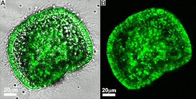
Fluorescence and bright-light images superimposed (B) Fluorescence image alone

Starting with fertilization, a Weizmann Institute-led team has traced the initial steps in the construction of the skeleton in live embryos – those of sea urchins. As reported recently in the Proceedings of the National Academy of Sciences, USA, they have gained surprising new insights into this elaborate process. 

 
A scanning electron microscope image showing a vesicle containing calcium carbonate nanospheres, 20 to 30 nanometers across, in a flash-frozen sea urchin embryo


To accumulate sufficient calcium and other minerals, each sea urchin embryo, which is about the size of a cross section of a human hair, needs to use all the calcium contained in hundreds of times its own volume of seawater. For decades, scientists had thought that the take-up of calcium and carbonate from the water and the actual building of the skeleton were performed exclusively by specialized embryonic cells. But in the new study, when the researchers observed the growth of the sea urchin embryo in sea water containing calcium ions labeled with green fluorescent dye, they were amazed to discover that the entire embryo was soon lit up in green: Evidently, many of its cells had absorbed the calcium ions.

To make sure the finding had not been produced accidentally, the scientists confirmed the wide distribution of the mineral using several advanced technologies: In addition to observing live embryos with a light laser microscope, they investigated frozen embryonic samples with a scanning electron microscope. Moreover, they used an innovative Israeli system – a multi-modal imaging station featuring fluorescence microscopy, elemental mapping and a scanning electron microscope that allows the examination of samples in open air rather than in a vacuum. The study was performed by Prof. Lia Addadi, Prof. Stephen Weiner and graduate student Netta Vidavsky, all of the Weizmann Institute’s Structural Biology Department, together with Weizmann Institute graduate Dr. Sefi Addadi of B-nano Ltd., Dr. Julia Mahamid, a Weizmann graduate currently in Germany, and Dr. Eyal Shimoni of Weizmann’s Chemical Research Support Department, as well as David Ben-Ezra and Prof. Muki Shpigel of the Israel Oceanographic and Limnological Research Institute in Eilat.    
 
(l-r) Dr. Eyal Shimoni, Dr. Sefi Addadi, Prof. Lia Addadi, Netta Vidavsky and Prof. Stephen Weiner
 
 
The study also revealed that when calcium carbonate gets into the embryo’s cells, it forms solid granules composed entirely of nanospheres. This texture is characteristic of many amorphous minerals and is known to be an intermediate stage in the formation of the skeletal tissue of the sea urchin, as revealed in studies by Weiner and Addadi more than a decade ago. The granules, in turn, were found to be stored in vesicles, spherical container-like structures.
 
A scanning electron microscope image of a flash-frozen sea urchin embryo, showing a large number of intra-cellular vesicles


These findings point to new avenues for studying skeleton formation in a wide range of living organisms, including humans. The fact that the entire embryo is mobilized for construction of the skeleton suggests that in humans, contrary to accepted belief, in addition to the specialized osteoblasts, other cells might be involved in the construction of bones and teeth. Moreover, the temporary storage of calcium carbonate in vesicles might be applicable to organisms other than sea urchins.

Elucidating these mechanisms is of enormous importance for understanding biological mineralization – an understanding that in turn could be crucial for future investigations into various diseases and structural abnormalities affecting bones and teeth.
 
 
Prof. Lia Addadi's research is supported by the Gerhardt M.J. Schmidt Minerva Center on Supramolecular Architecture; the Ilse Katz Institute for Material Sciences and Magnetic Resonance Research; and the Carolito Stiftung. Prof. Lia Addadi is the incumbent of the Dorothy and Patrick Gorman Professorial Chair.
 
Prof. Stephen Weiner's research is supported by the Helen and Martin Kimmel Center for Archaeological Science, which he heads; the J & R Center for Scientific Research; the Maurice and Vivienne Wohl Charitable Foundation; the Exilarch's Foundation; the estate of Hilda Jacoby-Schaerf; the estate of George and Beatrice F. Schwartzman; and the European Research Council. Prof. Weiner is the incumbent of the Dr. Walter and Dr. Trude Borchardt Professorial Chair in Structural Biology.


Life Sciences
English

Time is of the Essence

English
New findings in mice suggest that merely changing meal times could have a significant effect on the levels of triglycerides in the liver. The results of this Weizmann Institute of Science study, recently published in Cell Metabolism, not only have important implications for the potential treatment of metabolic diseases, they may also have broader implications for most research areas in the life sciences.

Many biological processes follow a set timetable, with levels of activity rising and dipping at certain times of the day. Such fluctuations, known as circadian rhythms, are driven by internal “body clocks” based on an approximately 24-hour period – synchronized to light-dark cycles and other cues in an organism’s environment. Disruption to this optimum timing system in both animal models and in humans can cause imbalances, leading to such diseases as obesity, metabolic syndrome and fatty liver. Night-shift workers, for example, have been shown to have higher incidence of these diseases. 
timing of meals (Image: Thinkstock)
In studying the role of circadian rhythm in the accumulation of lipids in the liver, postdoctoral fellow Yaarit Adamovich and the team in the lab of Dr. Gad Asher of the Weizmann Institute’s Biological Chemistry Department, together with scientists from Dr. Xianlin Han’s lab in the Sanford-Burnham Medical Research Institute, Orlando, US, quantified hundreds of different lipids present in the mouse liver. They discovered that a certain group of lipids, namely the triglycerides (TAG), exhibit circadian behavior, with levels peaking about eight hours after sunrise. The scientists were astonished to find, however, that daily fluctuations in this group of lipids persist even in mice lacking a functional biological clock, albeit with levels cresting at a completely different time – 12 hours later than the natural schedule. “These results came as a complete surprise: One would expect that if the inherent clock mechanism is ‘dead,’ TAG could not accumulate in a time-dependent fashion,” says Adamovich. So what was making the fluctuating lipid levels “tick” if not the clocks? “One thing that came to mind was that, since food is a major source of lipids – particularly TAG – the eating habits of these mice might play a role.” Usually, mice consume 20% of their food during the day and 80% at night. However, in mice lacking a functional clock, the team noted that they ingest food constantly throughout the day. This observation excluded the possibility that food is responsible for the fluctuating patterns seen in TAG levels in these mice. 

When the scientists proceeded to check the effect of an imposed feeding regimen upon wild type mice, however, they were in for another surprise: After they provided the same amount of food – but restricted 100% of the feeding to nighttime hours – the team observed a dramatic 50% decrease in overall liver TAG levels. 

These results suggest that the time at which TAG accumulation occurs, as well as its levels, are determined by the clocks together with timing of meals. The details of the mechanism that drives the actual fluctuating behavior are yet to be discovered. 

Asher: “The striking outcome of restricted nighttime feeding – lowering liver TAG levels in the very short time period of 10 days in the mice – is of clinical importance. Hyperlipidemia and hypertriglyceridemia are common diseases characterized by abnormally elevated levels of lipids in blood and liver cells, which lead to fatty liver and other metabolic diseases. Yet no currently available drugs have been shown to change lipid accumulation as efficiently and drastically as simply adjusting meal time – not to mention the possible side effects that may be associated with such drugs.” Of course, mice are nocturnal animals, so in order to construe these results for humans, the timetable would need to be reversed. 

Time is a crucial element in all biological systems, so these findings are likely to impact biological research in general: Circadian clock mechanisms function even in cultured cells, so research results could vary depending on the time at which samples are analyzed, or, with animals, their feeding regimen might significantly affect the experimental outcomes. In other words, when it comes to designing experiments, scientists should be aware that “timing is everything.”
 
 
Dr. Gad Asher’s research is supported by the Willner Family Leadership Institute; the Yeda-Sela Center for Basic Research; the Adelis Foundation; the Abisch Frenkel Foundation for the Promotion of Life Sciences; the Samuel M. Soref & Helene K. Soref Foundation; the late Rudolfine Steindling; and the Estate of Dorothy Geller.


timing of meals (Image: Thinkstock)
Life Sciences
English

Pages