We do not live by bread alone. To digest the bread, as well as the rest of the food we consume, our bodies resort to the help of microbes that live in our gut. These number in the billions; their total weight adds up to nearly two kilograms. Most are friendly and, as noted, even vital for the normal functioning of digestion and other body systems. But occasionally, disease-causing microbes such as Salmonella sneak in among them.
A team of Weizmann Institute scientists headed by
Dr. Guy Shakhar of the Immunology Department recently shed light on this mystery.
As reported in the journal
Immunity, the scientists showed that the immune system sends its “spies,” so-called dendritic cells, to the surface of the gut lining. The research was performed by graduate student Julia Farache, along with her lab-mates Idan Koren, Idan Milo and Dr. Irina Gurevich; Drs. Ki-Wook Kim and Ehud Zigmond from
Prof. Steffen Jung’s lab in the same department; and researchers from the Mt. Sinai School of Medicine in New York: Drs. Glaucia C. Furtado and Sergio A. Lira.
Using a two-photon microscope, the scientists created an innovative setup that enabled them to monitor in real time immune cells in the gut of a live mouse. It turns out that the moment Salmonella bacteria stick to the epithelium of the small intestine, the epithelial cells inform the immune system and, within half an hour, dendritic cells are recruited to the site of the infection. In video clips created under the microscope, these cells can be clearly seen squeezing through crowed tissue to reach the upper layer of the epithelium and sending their extensions – the dendrites for which they are named – to capture the bacteria.
Why do they respond in this manner to Salmonella but not to the millions of “good” bacteria in the same environment? While the beneficial bacteria don’t damage cells because they probably don’t stick to the gut lining, Salmonella signals its intent to harm by latching on to the epithelium.
Having swallowed the bacteria, the dendritic cells rush to report to the immune system. They start expressing receptors that guide them back into the intestinal tissue and through the lymph vessels. In the lymph nodes, they present fragments of Salmonella’s proteins – in other words, the bacterium’s “body parts” – to immune T cells, which turn on mechanisms that destroy the Salmonella, preventing poisoning.
This research may in the future help develop therapies against inflammatory bowel diseases, which are characterized by flare-ups of inflammation. Since dendritic cells are involved in igniting these flare-ups, possibly by overreacting to an infection, understanding their mechanism of action in the gut may help prevent their harmful activity.
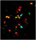
Dendritic cells send their extensions (red arrows) between adjacent epithelial cells (blue) in the lining of the small intestine
The new findings may also help develop oral vaccines, which offer major advantages over conventional methods of vaccination; for one, it’s easier to convince people to take a pill than to get an injection. Vaccines consist of weakened microbes, but for them to be effective, the microbes must be weakened in such a manner that they are capable of activating the immune system, yet do not cause disease yet. Therefore, it’s important to understand how microbes communicate with the immune system in the intestines, which is precisely what the current study has helped achieve.
Dendritic cells (brown) capture Salmonella microbes (light blue), then rapidly retract their extensions (arrow)
Prof. Steffen Jung's research is supported by the Leir Charitable Foundations; the Leona M. and Harry B. Helmsley Charitable Trust; the Adelis Foundation; Lord David Alliance, CBE; The Wolfson Family Charitable Trust; and the estate of Olga Klein Astrachan.
Dr. Guy Shakhar's research is supported by the Clore Center for Biological Physics; the Yeda-Sela Center for Basic Research; the Leona M. and Harry B. Helmsley Charitable Trust; the Dr. Dvora and Haim Teitelbaum Endowment Fund; Simone Pastor, Monaco; Lord David Alliance, CBE; Paul and Tina Gardner, Austin, TX; the Steven and Beverly Rubenstein Charitable Foundation; and the Paul Sparr Foundation.


, axons that lack KIF2A remain intact")


, and each column to a sample taken from a patient. Every colored spot stands for a number – the value of the “deregulation score” of the corresponding pathway, as determined for a particular patient. Dark blue stands for the activity of the pathway in normal brain tissue, whereas dark red indicates a high level of deviation from normal behavior. The clearly distinct group of normal brain samples appears as the dark blue vertical stripe, TgS7, in the middle. The TgS15 stripe corresponds to the newly identified subgroup of patients with longer survival prospects")

, and each column to a sample taken from a patient. Every colored spot stands for a number – the value of the “deregulation score” of the corresponding pathway, as determined for a particular patient. Dark blue stands for the activity of the pathway in normal brain tissue, whereas dark red indicates a high level of deviation from normal behavior. The clearly distinct group of normal brain samples appears as the dark blue vertical stripe, TgS7, in the middle. The TgS15 stripe corresponds to the newly identified subgroup of patients with longer survival prospects")


.jpg "Anti-donor immune T cells (green) attach themselves (blue arrows) to the donor’s veto cells (red); this binding leads to their destruction by the veto cells. Viewed under a two-photon microscope")
")


Alternate Endings
To save lives, it is sometimes vital to know as much as possible about death, in particular, the death of cells. For example, cancer chemotherapy works by activating a cellular death program called apoptosis. But if the molecular machinery of apoptosis is defective, or if the cancer cells learn to avoid apoptosis, which indeed they often do, chemotherapy becomes ineffective. Therefore, the recent discovery at the Weizmann Institute of so-called germ cell death – an alternative cell death pathway – can be of great value for the development of future life-saving therapies.
In short
• Some germ cells – sperm precursors – undergo a special form of cell death that ensures quality control.
• Germ cell death differs from the main type – apoptosis – in a number of crucial ways.
• These may point to new mechanisms for anti-cancer therapies.
Sperm cells are formed in a seemingly wasteful manner, which probably serves to ensure quality control: Germ cells are constantly created in large numbers, after which many of them die. By tracking the death of those cells in a living organism, the researchers were able to reveal its mechanics in great detail.
The researchers discovered that a central role in germ cell death is played by the mitochondria: These organelles activate a particular gene, htrA2, which makes a destructive protease enzyme. HtrA2 has an equivalent in organisms ranging from bacteria to mammals, which suggests that the findings of the Weizmann fruit fly study are applicable to humans. Yet another major component of the germ cell death mechanism is the lysosome, the cell’s stomach-like organelle that is filled with enzymes for breaking down cellular waste and debris. Lysosomes contribute to cellular destruction by spilling out their contents.