The Spies Inside

English

Dr. Guy Shakhar


We do not live by bread alone. To digest the bread, as well as the rest of the food we consume, our bodies resort to the help of microbes that live in our gut. These number in the billions; their total weight adds up to nearly two kilograms. Most are friendly and, as noted, even vital for the normal functioning of digestion and other body systems. But occasionally, disease-causing microbes such as Salmonella sneak in among them.

In most cases, the immune system identifies and destroys the dangerous microbes so that we do not even know we’d been exposed to the risk. But how does the system tell the good microbes from the bad? How does it detect the danger? The task is far from trivial: The microbes are contained in the gut, whereas the immune cells are embedded in the gut lining, the epithelium. Complicating detection even further, the few harmful bacteria present in a healthy gut are vastly outnumbered by the rest of the gut flora.
 
 
Live two-photon microscopy of the small intestine
 
A team of Weizmann Institute scientists headed by Dr. Guy Shakhar of the Immunology Department recently shed light on this mystery. As reported in the journal Immunity, the scientists showed that the immune system sends its “spies,” so-called dendritic cells, to the surface of the gut lining. The research was performed by graduate student Julia Farache, along with her lab-mates Idan Koren, Idan Milo and Dr. Irina Gurevich; Drs. Ki-Wook Kim and Ehud Zigmond from Prof. Steffen Jung’s lab in the same department; and researchers from the Mt. Sinai School of Medicine in New York: Drs. Glaucia C. Furtado and Sergio A. Lira.

Using a two-photon microscope, the scientists created an innovative setup that enabled them to monitor in real time immune cells in the gut of a live mouse. It turns out that the moment Salmonella bacteria stick to the epithelium of the small intestine, the epithelial cells inform the immune system and, within half an hour, dendritic cells are recruited to the site of the infection. In video clips created under the microscope, these cells can be clearly seen squeezing through crowed tissue to reach the upper layer of the epithelium and sending their extensions – the dendrites for which they are named – to capture the bacteria.

Why do they respond in this manner to Salmonella but not to the millions of “good” bacteria in the same environment? While the beneficial bacteria don’t damage cells because they probably don’t stick to the gut lining, Salmonella signals its intent to harm by latching on to the epithelium.

Having swallowed the bacteria, the dendritic cells rush to report to the immune system. They start expressing receptors that guide them back into the intestinal tissue and through the lymph vessels. In the lymph nodes, they present fragments of Salmonella’s proteins – in other words, the bacterium’s “body parts” – to immune T cells, which turn on mechanisms that destroy the Salmonella, preventing poisoning.

This research may in the future help develop therapies against inflammatory bowel diseases, which are characterized by flare-ups of inflammation. Since dendritic cells are involved in igniting these flare-ups, possibly by overreacting to an infection, understanding their mechanism of action in the gut may help prevent their harmful activity.
 
Dendritic cells send their extensions (red arrows) between adjacent epithelial cells (blue) in the lining of the small intestine

The new findings may also help develop oral vaccines, which offer major advantages over conventional methods of vaccination; for one, it’s easier to convince people to take a pill than to get an injection. Vaccines consist of weakened microbes, but for them to be effective, the microbes must be weakened in such a manner that they are capable of activating the immune system, yet do not cause disease yet. Therefore, it’s important to understand how microbes communicate with the immune system in the intestines, which is precisely what the current study has helped achieve.
 
Dendritic cells (brown) capture Salmonella microbes (light blue), then rapidly retract their extensions (arrow)
 
Prof. Steffen Jung's research is supported by the Leir Charitable Foundations; the Leona M. and Harry B. Helmsley Charitable Trust; the Adelis Foundation; Lord David Alliance, CBE; The Wolfson Family Charitable Trust; and the estate of Olga Klein Astrachan.
 
Dr. Guy Shakhar's research is supported by the Clore Center for Biological Physics; the Yeda-Sela Center for Basic Research; the Leona M. and Harry B. Helmsley Charitable Trust; the Dr. Dvora and Haim Teitelbaum Endowment Fund; Simone Pastor, Monaco; Lord David Alliance, CBE; Paul and Tina Gardner, Austin, TX; the Steven and Beverly Rubenstein Charitable Foundation; and the Paul Sparr Foundation.


Live two-photon microscopy of the small intestine
Life Sciences
English

Veto on Disease

English


Prof. Yair Reisner


A little tolerance can go a long way. If the immune system, for instance, could be coaxed into tolerating foreign tissue rather than rejecting it outright, a variety of blood disorders could be treated by bone marrow transplants. Immune tolerance could also help extend these transplants to people who are currently too frail to receive them; in addition, it could facilitate the transplantation of kidneys, liver and other body organs.

An international team led by Weizmann Institute researchers has recently managed to induce a certain degree of immune tolerance by making use of so-called veto cells. These are naturally occurring immune cells that exert veto power over their own destruction: When attacked by the recipient’s immune cells, they launch a pre-emptive strike, destroying the attackers.

Lead study author Prof. Yair Reisner of the Institute’s Immunology Department  has been studying veto cells for more than a decade, focusing on certain types known as CD8+. These cells exhibit the most potent veto activity of all, but they may be toxic to the patient because they can induce a complication known as graft-versus-host disease, in which the transplant attacks the host. Reisner’s team has found a way of circumventing this problem by generating a line of less toxic CD8+ veto cells. In the new study, reported in Blood, he and colleagues have now prepared the ground for using these cells in patients.  According to the accompanying editorial entitled “Location, Location, Location,” the study has revealed the conditions in which the “promise of veto cells, as a cellular therapy, may finally be realized.”

As suggested in the editorial’s title, the scientists have determined the exact location in which veto cells exert their protective effect: the lymph nodes. The researchers have also revealed the mechanism of the cells’ action. It turns out that the cells cast their veto as soon as the recipient’s immune system T cells, which continuously patrol the body looking for foreign invaders, recognize the newly transplanted veto cells and latch onto them. That is precisely when the veto cells make their preemptive move: Equipped with ready-made poisons, they destroy the attackers, whose own poisons take longer to be produced. These details were revealed in collaboration with the lab of Dr. Guy Shakhar, also of the Immunology Department, thanks to an innovative two-photon microscopy system that made it possible to observe individual immune cells in a live mouse in real time.

The scientists further determined how to make sure veto cells reach their destination: Prior to transplantation, they must be grown in culture with the help of the biochemical interleukin-15, which preserves the “homing” receptor that guides them to the lymph nodes.
 
 
Anti-donor immune T cells (green) attach themselves (blue arrows) to the donor’s veto cells (red); this binding leads to their destruction by the veto cells. Viewed under a two-photon microscope

 
Indeed, in the study, veto cells successfully eliminated they type of recipient T cells that are responsible for much of the rejection. As a result, white mice tolerated skin grafts from black mice. Normally, such transplants require a drastic suppression of the recipient’s immune system with drugs and radiation to prevent rejection, but thanks to the tolerance induced by veto cells, the white mice did not reject the black patches even though their immune systems had been only mildly suppressed. Further research is needed to establish whether this approach can be as effective in humans as it is in laboratory animals.

Major and equal contributions to this study, conducted in Reisner’s lab, were made by graduate students Dr. Eran Ophir and Noga Or-Geva. In an earlier, preliminary study in Reisner’s lab, led by Ophir and Dr. Yaki Edelstein and published in Blood in 2010,  the concept of using veto cells to induce tolerance had been established in a more artificial mouse model for bone marrow graft rejection.  

Currently, bone marrow transplants are used only to treat disorders that are otherwise fatal because the treatment itself is risky. The major risk is infection, which stems from the suppression of the immune system in preparation for the transplant. But if the suppression can be mild, as was the case in the current study, bone marrow transplants could potentially be extended to such nonfatal disorders as sickle-cell anemia and thalassemia. They could also be made available to people who are presently unable to benefit from such transplants because they are too vulnerable to undergo a massive suppression of the immune system, such as elderly patients with leukemia or lymphoma. Moreover, the veto cell approach could be used to induce tolerance toward transplanted kidneys or other organs without the need for continuous immune suppression.
 
Prof. Yair Reisner's research is supported by the Belle S. and Irving E. Meller Center for the Biology of Aging, which he heads; the Leona M. and Harry B. Helmsley Charitable Trust; the Steven and Beverly Rubenstein Charitable Foundation; Roberto and Renata Ruhman, Brazil; the wstate of Samuel and Valerie Rodetsky; and the Estate of Lola Asseof. Prof. Reisner is the incumbent of the Henry H. Drake Professorial Chair in Immunology.
 
 
Life Sciences
English

Bringing Down Barriers

English
 
 
Blood brain barrier. Image: Ben Brahim Mohammed via Wikimedia Commons


Our brains are well protected, and for a good reason. Most of the proteins present at high levels in the blood, such as antibodies or the regulatory molecule albumin, are kept out of the brain because they could damage it or disturb its exquisitely organized functioning. Only essential nutrients are allowed entry from the circulation.
Prof. Matityahu Fridkin


This tight control is imposed by the blood-brain barrier. The barrier is both physical – the cells lining the blood capillaries in the brain are tightly attached to one another – and chemical: The brain’s lining chemically repels potentially damaging substances. When essential nutrients are needed, the barrier creates “openings” called physiological gates – chemical and physical changes in the lining that permit these nutrients to enter.
 
Although this barrier is vital for health, it poses a major problem for treating brain disorders. For instance, since most drugs cannot get across this barrier, malignant brain tumors cannot be treated efficiently by regular anti-cancer chemotherapy. Scientists have attempted to overcome this problem by “smuggling” drugs into the brain with the help of “shuttle” proteins capable of passing through physiological gates. But unfortunately, these gates are relatively few in number, so that insufficient quantities of the drug are delivered to the brain using this strategy.
 
A potential solution might come from an unexpected direction: the study of HIV, the virus that causes AIDS. HIV is a master of penetration: It gets into mammalian cells – particularly immune T cells – with great ease and can even migrate from cell to cell. Not only that, in 1996, scientists discovered that HIV can cross the blood-brain barrier, penetrating the central nervous system – the brain and spinal cord. This was a surprising discovery because the two receptors known to help the virus infect T cells are not found on cells lining the brain’s protective barrier. The mystery was later resolved when the scientists found that the virus manufactures its own “opener” for the barrier: a molecule called HIV-1-TAT-protein, which contains unusual sequences of amino acids in two of its regions. Even when purified from the virus, the HIV-1-TAT-protein maintained its barrier-opening capacity.
Prof. Yoram Shechter


It is precisely this capacity that drew the attention of Weizmann Institute scientists. Prof. Yoram Shechter of the Biological Chemistry Department, Prof. Matityahu Fridkin of the Organic Chemistry Department in the Chemistry Faculty and Dr. Itzik Cooper of the Neurobiology Department decided to test the possibility that the blood-brain barrier could be opened by short peptides based on the unusual sequence of amino acids in the HIV-1-TAT protein.
 
To perform their test, they used an experimental system developed in the laboratory of the late Prof. Vivian Teichberg of Weizmann’s Neurobiology Department, which uniquely simulates the blood-brain barrier in a laboratory dish. This system consists of a layer of tightly linked cells drawn from the lining of pig brains and grown in culture. The tightness of the layer is measured by an evaluation of its electrical resistance. When this tightness drops, the permeability of the layer is further quantified with the help of radioactively labeled proteins.
Dr. Itzik Cooper
 
The scientists found that their HIV-1-TAT peptides were indeed able to weaken the blood-brain barrier, permitting the entry of therapeutic proteins and other drugs. Most important, as reported recently in the Journal of Biological Chemistry, the drugs penetrated the barrier in quantities sufficient for the treatment of major brain disorders. Further studies are being conducted at the Weizmann Institute in collaboration with Prof. Yosef Yarden of Weizmann’s  Biological Regulation Department and Prof. Yael Mardor of the Chaim Sheba Medical Center at Tel Hashomer.

Obviously, destabilizing the blood-brain barrier is undesirable in healthy people, but in cancer patients this approach could represent the least of all evils. In fact, in certain non-fatal diseases, this barrier is markedly disrupted, suggesting that such a disruption poses no immediate threat to a person’s life. The new Weizmann Institute approach could therefore be valid for treating brain tumors and other neurological disorders.
 
Prof. Matityahu Fridkin’s research is supported by the Adelis Foundation.

Prof. Yoram Shechter’s research is supported by the Adelis Foundation.


Life Sciences
English

A Question of Upbringing

English
 
 
The behavior (gene expression) 
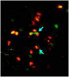
of immune cells in the colon (l-r): 1) monocytes before arrival in the colon; 2) “delinquent” uneducated cells; 3) “educated” immune cells; 4) resident immune cells


Failure to educate the young generation properly can have devastating consequences – not only among human beings, but among cells too. Uneducated immune cells, it turns out, might be at least partly responsible for the notorious flare-ups that occasionally occur in chronic inflammatory diseases of the digestive tract.

Our digestive tract is teeming with bacteria – some inhabiting it permanently and assisting in digestion, others introduced with food and drink. Keeping such a dynamic system in balance requires ongoing maintenance. A central role in this process is played by immune cells called macrophages, or "big eaters," believed to play an important role in clearing debris and keeping tissues healthy. Macrophages are generated from precursor cells, the monocytes, which arrive from the blood and settle in the lining of the gut.  

A collaborative study led by Weizmann Institute researchers has revealed that for about ten days, all newly recruited monocytes are taught – probably by signals originating in the gut lining – to express the same genes as do the more veteran immune cells. Once their education is complete, these cells start dutifully contributing to the proper maintenance of the lining.
 
 
(l-r) Prof. Steffen Jung, and Drs. Ehud Zigmond and Chen Varol
 
But if the gut lining is already inflamed – for example, as a result of bacterial infection – the newly arrived monocytes display a strikingly different behavior. Like teenagers giving in to peer pressure, these cells, on landing in a bad environment, turn bad themselves. They fail to undergo the proper education, so that their gene expression follows an abnormal pattern. As a result, instead of helping to maintain the gut, they start promoting inflammation, making it even worse than it was when they arrived.

Such misbehaving immune cells might help explain what happens during flare-ups of inflammatory bowel disease: Even a slight disruption resulting from exposure to bacteria or certain foods, which would normally be quickly corrected in a healthy person, leads to lasting inflammation. And once such inflammation is in place, the newly arriving monocytes, acting like juvenile delinquents who refuse to be educated, apparently keep aggravating this inflammation instead of correcting it.

The study, reported recently in Immunity, was led by Prof. Steffen Jung of the Weizmann Institute’s Immunology Department and performed in transgenic mice developed in his laboratory. The research was conducted by Dr. Ehud Zigmond, a physician and Ph.D. student, and Dr. Chen Varol of the Tel Aviv Sourasky Medical Center, in collaboration with departmental colleagues Dr. Guy Shakhar and Julia Farache, as well as Dr. Gilgi Friedlander of  Biological Services. Reagents were provided by Dr.  Kenneth M. Murphy of Washington University School of Medicine, Dr. Matthias Mack of the University of Regensburg in Germany, Dr. Nahum Shpigel of the Hebrew University of Jerusalem and Dr. Ivo G. Boneca of the Pasteur Institute and INSERM in France.

The findings point to new ways of treating inflammatory bowel disease. One approach would be to temporarily interrupt the recruitment of new monocytes to the gut once inflammation has begun, to prevent its further aggravation. In their studies in mice, Jung and his team indeed showed that antibodies blocking the arrival of new monocytes to the colon alleviated the symptoms of inflammation.

But in the longer term, as in many other areas of life, it is education that holds the real promise for the future. Once scientists understand in greater detail how monocyte education takes place in the gut lining, they should be able to ensure that it runs its proper course, so that the monocyte “kids” behave themselves, contributing to health rather than disease.
 
Prof.Steffen Jung’s research is supported by the Jeanne and Joseph Nissim Foundation for Life Sciences Research; the Leir Charitable Foundations; the Leona M. and Harry B. Helmsley Charitable Trust; the Adelis Foundation; Lord David Alliance, CBE; the Wolfson Family Charitable Trust; the estate of Olga Klein Astrachan; and the estate of Florence Cuevas.

Dr. Guy Shakhar’s research is supported by the Clore Center for Biological Physics; the Yeda-Sela Center for Basic Research; the Jeanne and Joseph Nissim Foundation for Life Sciences Research; the Leona M. and Harry B. Helmsley Charitable Trust; the Dr. Dvora and Haim Teitelbaum Endowment Fund; Simone Pastor, Monaco; Lord David Alliance, CBE; Paul and Tina Gardner, Austin, TX; the Steven and Beverly Rubenstein Charitable Foundation; and the Paul Sparr Foundation.


Life Sciences
English

How Stem Cells Stay Young

English
 
Hiding deep inside the bone marrow, special cells wait patiently for the hour of need – infection, for example – at which point these blood-forming stem cells can proliferate and differentiate into billions of mature blood immune cells. But the body always maintains a reserve of undifferentiated stem cells for future crises. A research team headed by Prof. Tsvee Lapidot of the Institute’s immunology Department recently discovered a new type of bodyguard that protects stem cells from over-differentiation. In a paper that appeared in Nature Immunology, they revealed how this rare, previously unknown sub-group of activated immune cells keeps the stem cells in the bone marrow “forever young.”

Blood-forming stem cells live in comfort in the bone marrow, surrounded by an entourage of support cells that cater to their needs and direct their development – the mesenchymal cells. The research team, which included postdoctoral fellow Dr. Aya Ludin, Prof. Steffen Jung of the Immunology Department and his group, and Ziv Porat of the Biological Services Unit, discovered another type of support cell for the stem cells. These cells are an offshoot of the macrophage family, literally the “big eaters” of the immune system, which are important, for instance, for bacterial clearance. It is a rare sub-population of the bone-marrow macrophages that take stem cells under their wing and prevent differentiation.
 
Prostaglandins guard stem cells by increasing the production of an inhibitory factor in the mesenchymal bone marrow cells. Mesenchymal cells are marked with a green protein (left); the inhibitory factor is red (center). Combined image (right): Treatment with prostaglandins (bottom) increases the secretion of the inhibitory factor


The researchers revealed, in precise detail, how these macrophages guard the stem cells. They secrete substances called prostaglandins, which are absorbed by the stem cells. In a chain of biochemical events, these substances delay differentiation and preserve the youthful state of the stem cells. In addition, the prostaglandins work on the neighboring mesenchymal cells, activating the secretion of a delaying substance in them and increasing the production of receptors for this substance on the stem cells themselves. This activity, says Lapidot, may be what helps the non-dividing stem cells survive chemotherapy – a known phenomenon. Macrophages also live through the treatment, and they respond by increasing their prostaglandin output, thus heightening their vigilance in protecting the stem cells.
 
In previous work in Lapidot’s lab, it was discovered that prostaglandin treatments can improve the number and quality of stem cells. This insight is currently being tested by doctors in clinical trials for the use of stem cells transplanted from umbilical cord blood to treat adult leukemia patients. These trials are showing that prior treatment with prostaglandins improves the migration and repopulation potential, so that the small quantities of stem cells in cord blood can better cure the patients.
 
Prof. Steffen Jung's research is supported by the Jeanne and Joseph Nissim Foundation for Life Sciences Research; the Leir Charitable Foundations; the Leona M. and Harry B. Helmsley Charitable Trust; the Adelis Foundation; Lord David Alliance, CBE; the Wolfson Family Charitable Trust; the estate of Olga Klein Astrachan and the estate of Florence Cuevas.
 
Prof. Tsvee Lapidot's research is supported by the M.D. Moross Institute for Cancer Research; and the Leona M. and Harry B. Helmsley Charitable Trust. Prof. Lapidot is the incumbent of the Edith Arnoff Stein Professorial Chair in Stem Cell Research.


Prostaglandins guard stem cells by increasing the production of an inhibitory factor in the mesenchymal bone marrow cells. Mesenchymal cells are marked with a green protein (left); the inhibitory factor is red (center). Combined image (right): Treatment with prostaglandins (bottom) increases the secretion of the inhibitory factor
Life Sciences
English

Stem Cell Bodyguards

English


Hiding deep inside the bone marrow are special cells. They wait patiently for the hour of need, at which point these blood-forming stem cells can proliferate and differentiate into billions of mature blood immune cells to help the body cope with infection, for example, or extra red blood cells for low oxygen levels at high altitudes. Even in emergencies, however, the body keeps to a long-term plan: It maintains a reserve of undifferentiated stem cells for future needs and crises. A research team headed by Prof. Tsvee Lapidot of the Institute’s immunology Department recently discovered a new type of bodyguard that protects stem cells from over-differentiation. In a paper that appeared in Nature Immunology, they revealed how this rare, previously unknown sub-group of activated immune cells keeps the stem cells in the bone marrow “forever young.”

Blood-forming stem cells live in comfort in the bone marrow, surrounded by an entourage of support cells that cater to their needs and direct their development – the mesenchymal cells. But the research team, which included postdoctoral fellow Dr. Aya Ludin, Prof. Steffen Jung of the Immunology Department and his group, and Ziv Porat of the Biological Services Unit, discovered another type of support cell for the stem cells. These are an offshoot of the macrophage family – literally the “big eaters” of the immune system that are important, for instance, for bacterial clearance. The team found that a rare sub-population of the bone-marrow macrophages has another role to play. Each of these rare macrophages can take a stem cell under its wing and prevent its differentiation.

Probing more deeply, the researchers revealed, in precise detail, how these macrophages guard the stem cells. They secrete substances called prostaglandins, which are absorbed by the stem cells. In a chain of biochemical events, these substances delay differentiation and preserve the youthful state of the stem cells. In addition, the prostaglandins work on the neighboring mesenchymal cells, activating the secretion of a delaying substance in them and increasing the production of receptors for this substance on the stem cells, themselves. This activity, says Lapidot, may help the non-dividing stem cells survive chemotherapy – a known phenomenon. Macrophages also live through the treatment, and they respond by increasing their prostaglandin output, thus heightening their vigilance in protecting the stem cells.

The bodyguard macrophages also increase their activity in times of infection. While other members of the macrophage family are recruited to fight the pathogens, their cousins in the bone marrow are hard at work ensuring that a pool of stem cells will resist the urge to differentiate.

In previous work in Lapidot’s lab, it was discovered that prostaglandin treatments can improve the number and quality of stem cells. This insight is currently being tested by doctors in clinical transplantation trials for the use of stem cells from umbilical cord blood to treat adult leukemia patients. These trials are showing that prior treatment with prostaglandins improves migration and repopulation potential, enabling the small quantities of cord blood stem cells to better cure the patients. “The present study hints at the possibility of further increasing the support for bone marrow stem cells by exploring this intriguing connection between the immune cells and stem cells,” says Lapidot. “An understanding of the mechanisms at work in these cells might improve the success of stem cell transplantation, especially that of umbilical blood.”
 
Prostaglandins guard stem cells by increasing the production of an inhibitory factor in the mesenchymal bone marrow cells. Mesenchymal cells are marked with a green protein (left); the inhibitory factor is red (center). Combined image (right): Treatment with prostaglandins (bottom) increases the secretion of the inhibitory factor
 
 
Prof. Steffen Jung’s research is supported by the Leir Charitable Foundations; the Leona M. and Harry B. Helmsley Charitable Trust; the estate of Olga Klein – Astrachan; the estate of Lola Asseof; Lord David Alliance, CBE; and the Adelis Foundation.

Prof. Tsvee Lapidot’s research is supported by the Leona M. and Harry B. Helmsley Charitable Trust. Prof. Lapidot is the incumbent of the Edith Arnoff Stein Professorial Chair in Stem Cell Research.


Prostaglandins guard stem cells by increasing the production of an inhibitory factor in the mesenchymal bone marrow cells. Mesenchymal cells are marked with a green protein (left); the inhibitory factor is red (center). Combined image (right): Treatment with prostaglandins (bottom) increases the secretion of the inhibitory factor
Life Sciences
English

Immune System Tricks

English
 

Prof. Irit SagiA team of Weizmann Institute scientists has turned the tables on an autoimmune disease. In such diseases,

including Crohn’s and rheumatoid arthritis, the immune system mistakenly attacks the body’s tissues. But the
scientists managed to trick the immune systems of mice into targeting one of the body’s players in autoimmune processes, an enzyme known as MMP9. The results of their research appeared in Nature Medicine.

Prof. Irit Sagi of the Biological Regulation Department and her research group have spent years looking for ways to home in on and block members of the matrix metalloproteinase (MMP) enzyme family. These proteins cut through the tough support materials in our bodies, which makes them crucial for cellular mobilization, proliferation and wound healing, among other things. But when the activities of some members of the MMP family, especially MMP9, get out of control, it can aid and abet autoimmune disease and cancer metastasis. Blocking these proteins, then, might lead to effective treatments for a number of diseases.   

Originally, Sagi and others had designed synthetic drug molecules to directly target MMPs. But these drugs proved to be fairly crude tools that had extremely severe side effects. The body normally produces its own MMP inhibitors, known as TIMPs, as part of the tight regulation program that keeps these enzymes in line. As opposed to the synthetic drugs, these work in a highly selective manner. An arm on each TIMP is precisely constructed to reach into a cleft in the enzyme that shelters the active bit – a metal zinc ion surrounded by three histidine peptides – closing it off like a snug cork. “Unfortunately,” says Sagi, “it is quite difficult to reproduce this precision synthetically.”

Dr. Netta Sela-Passwell began working on an alternative approach as an M.Sc. student in Sagi’s lab and continued on through her Ph.D. research. She and Sagi decided that, rather than attempting to design a synthetic molecule to directly attack MMPs, they would try to trick the immune system into creating natural antibodies that would target MMP9 through immunization. Just as immunization with a killed virus induces the immune system to create antibodies that then attack live viruses, an MMP immunization would get the body to make antibodies that would block the enzyme at its active site.

Together with Prof. Abraham Shanzer of the Organic Chemistry Department, they created an artificial version of the metal zinc-histidine complex at the heart of the MMP9 active site. They then injected these small, synthetic molecules into mice and afterward checked the mice’s blood for signs of immune activity against the MMPs. Indeed, the team identified antibodies, which they dubbed “metallobodies,” that were similar but not identical to TIMPS. A detailed analysis of their atomic structure suggested the two work in a similar way – reaching into the enzyme’s cleft and blocking the active site. The metallobodies were selective for just two members of the MMP family – MMP2 and 9 – and they bound tightly to the mouse versions of these enzymes, as well as to the human ones.
 
Left: Natural control mechanism blocks the enzyme's zinc active site. Right: Novel antibody works as effectively as the natural control mechanism


As the team hoped, when they had induced an inflammatory condition that mimics Crohn’s disease in mice, the symptoms were prevented when mice were treated with metallobodies. “We are excited not only by the potential of this method to possibly treat Crohn’s,” says Sagi, “but by the potential of using this approach to explore novel treatments for many other diseases.” Yeda, the technology transfer arm of the Weizmann Institute, has applied for a patent for the synthetic immunization molecules as well as the generated metallobodies.

Also participating in this research were Drs. Orly Dym, Haim Rozenberg, Rina Arad-Yellin and Tsipi Shoham, and Raanan Margalit of the Structural Biology, Immunology and Biological Regulation Departments; Raghavendra Kikkeri of the Organic Chemistry Department; Miriam Eisenstein of the Chemical Research Support Department; Ori Brenner of the Veterinary Resources Department; and Tamar Danon of the Molecular Cell Biology Department.
 
Prof. Irit Sagi's research is supported by the Spencer Charitable Fund; the Leona M. and Harry B. Helmsley Charitable Trust; Cynthia Adelson, Canada; Dr. Mireille Steinberg, Canada; the Leonard and Carol Berall Post Doctoral Fellowship; and the Ilse Katz Institute for Material Sciences and Magnetic Resonance Research. Prof. Sagi is the incumbent of the Maurizio Pontecorvo Professorial Chair.
 
Prof. Abraham Shanzer is the incumbent of the Siegfried and Irma Ullmann Professorial Chair.


Left: Natural control mechanism blocks the enzyme's zinc active site. Right: Novel antibody works as effectively as the natural control mechanism
Life Sciences
English

Phase III Clinical Trials of Diabetes Treatment

English
 
 
A treatment for Type 1 diabetes developed by Prof. Irun Cohen of the Institute’s Immunology Department has met both the primary and secondary goals of phase III clinical trials. Andromeda Biotech, which is licensed by Yeda Research and Development, Ltd. (the technology transfer arm of the Weizmann Institute) to develop the treatment, reported that it had been tested on 457 patients who had been diagnosed with Type 1 diabetes a short time before the trial. From the initial results, those who received the treatment – DiaPep277® – in the double-blinded test appeared to have significantly higher pancreas function than those in the control group. The trial took place over a period of two years, during which one group received DiaPep277® injections every three months and the control group was given a placebo. All received insulin, as needed, to control glucose levels.
Prof. Irun Cohen, DiaPep 227 developer
 
DiaPep277® is a unique peptide derived from the sequence of the human heat shock protein 60 (Hsp60). The peptide acts by modulating the immune system, preventing the destruction of the pancreatic cells that secrete insulin and preserving their natural function. Treatment of Type 1 diabetes patients with DiaPep277® may have several medical benefits: slowing the deterioration of the diseased tissue, improving metabolic control and reducing daily insulin requirements as well as the complications of diabetes.

The research and development team at Andromeda Biotech are currently working on a full assessment of the efficacy and safety data, and they are planning to conduct another clinical trial later this year.
 
Prof. Irun Cohen’s research is supported by the Laszlo N Tauber Family Foundation. 
 
 
Prof. Irun Cohen, DiaPep 227 developer
Life Sciences
English

New Antibodies Treat Autoimmune Disease in Mice

English
A team of Weizmann Institute scientists has turned the tables on an autoimmune disease. In such diseases, including Crohn’s and rheumatoid arthritis, the immune system mistakenly attacks the body’s tissues. But the scientists managed to trick the immune systems of mice into targeting one of the body’s players in autoimmune processes, an enzyme known as MMP9. The results of their research appear today in Nature Medicine.

Prof. Irit Sagi of the Biological Regulation Department and her research group have spent years looking for ways to home in on and block members of the matrix metalloproteinase (MMP) enzyme family. These proteins cut through such support materials in our bodies as collagen, which makes them crucial for cellular mobilization, proliferation and wound healing, among other things. But when some members of the family, especially MMP9, get out of control, they can aid and abet autoimmune disease and cancer metastasis. Blocking these proteins might lead to effective treatments for a number of diseases. 
 
Originally, Sagi and others had designed synthetic drug molecules to directly target MMPs. But these drugs proved to be fairly crude tools that had extremely severe side effects. The body normally produces its own MMP inhibitors, known as TIMPs, as part of the tight regulation program that keeps these enzymes in line. As opposed to the synthetic drugs, these work in a highly selective manner. An arm on each TIMP is precisely constructed to reach into a cleft in the enzyme that shelters the active bit – a metal zinc ion surrounded by three histidine peptides – closing it off like a snug cork. “Unfortunately,” says Sagi, “it is quite difficult to reproduce this precision synthetically.”

Dr. Netta Sela-Passwell began working on an alternative approach as an M.Sc. student in Sagi’s lab, and continued on through her Ph.D. research. She and Sagi decided that, rather than attempting to design a synthetic molecule to directly attack MMPs, they would try coaxing the immune system into targeting MMP-9 through immunization. Just as immunization with a killed virus induces the immune system to create antibodies that then attack live viruses, an MMP immunization would trick the body into creating antibodies that block the enzyme at its active site.

Together with Prof. Abraham Shanzer of the Organic Chemistry Department, they created an artificial version of the metal zinc-histidine complex at the heart of the MMP9 active site. They then injected these small, synthetic molecules into mice and afterward checked the mice’s blood for signs of immune activity against the MMPs. The antibodies they found, which they dubbed “metallobodies,” were similar but not identical to TIMPS, and a detailed analysis of their atomic structure suggested they work in a similar way – reaching into the enzyme’s cleft and blocking the active site. The metallobodies were selective for just two members of the MMP family – MMP2 and 9 – and they bound tightly to both the mouse versions of these enzymes and the human ones.

As they hoped, when they had induced an inflammatory condition that mimics Crohn's disease in mice, the symptoms were prevented when mice were treated with metallobodies. “We are excited not only by the potential of this method to treat Crohn’s,” says Sagi, but by the potential of using this approach to explore novel treatments for many other diseases.” Yeda, the technology transfer arm of the Weizmann Institute has applied for a patent for the synthetic immunization molecules as well as the generated metallobodies.

Also participating  in this research were Drs. Orly Dym, Haim Rozenberg, Rina Arad-Yellin and  Tsipi Shoham, and Raanan Margalit of the Structural Biology, Immunology and Biological Regulation Departments, Raghavendra Kikkeri of the Organic Chemistry Department, Miriam Eisenstein of the Chemical Research Support Department, Ori Brenner of the Veterinary Resources Department and Tamar Danon of the Molecular Cell Biology Department.
 
Prof. Irit Sagi’s research is supported by the Spencer Charitable Fund; the Leona M. and Harry B. Helmsley Charitable Trust; Cynthia Adelson, Canada; Mireille Steinberg, Canada; the Leonard and Carol Berall Post Doctoral Fellowship; and the Ilse Katz Institute for Material Sciences and Magnetic Resonance Research. Prof. Sagi is the incumbent of the Maurizio Pontecorvo Professorial Chair. 
 
This work was financially supported by Merck Serono S.A., a division of Merck KGaA, Darmstadt, Germany.
 

Left: Natural control mechanism blocks the enzyme's zinc active site. Right: Novel antibody works as effectively as the natural control mechanism

 
Left: Natural control mechanism blocks the enzyme's zinc active site. Right: Novel antibody works as effectively as the natural control mechanism
Life Sciences
English

Hide and Seek Signals

English
 
The white blood cells that fight disease and help our bodies heal are directed to sites of infection or injury by “exit signs” – chemical signals that tell them where to pass through the blood vessel walls and into the underlying tissue. New research at the Weizmann Institute, which appeared in Nature Immunology online, shows how the cells lining blood vessel walls may act as “selectors” by hiding the signals where only certain “educated” white blood cells will find them.

In previous studies, Prof. Ronen Alon and his team in the Immunology Department had found that near sites of inflammation, white blood cells rapidly crawl along the inner lining of the blood vessels with tens of tiny legs that grip the surface tightly, feeling for the exit sign. Such signs consist of migration-promoting molecules called chemokines, which the cells lining the blood vessels – endothelial cells – display on their outer surfaces like flashing lights.

In the new study, Alon and his team, including Drs. Ziv Shulman and Shmuel Cohen, found that not all chemokine signals produced by endothelial cells are on display. They observed the recruitment of subsets of immune cells called effector cells that are the “special forces” of the immune system: They receive training in the lymph nodes, where they learn to identify a particular newly-invading pathogen and then return to the bloodstream on a search and destroy mission. Like the other white blood cells, effector cells crawled on tiny appendages along the lining of inflamed blood vessels near the site of pathogen entry, but rather than sensing surface chemokines, they used their legs to reach into the endothelial cells in search of the migration-promoting chemokines.

As opposed to the external exit signs, these chemokines were held in tiny containers – vesicles – inside the inflamed endothelial cell walls. The effector cells paused in the joins where several cells met, inserting their legs through the walls of several endothelial cells at once to trap chemokines as they were released from vesicles at the endothelial cell membrane. Once they obtained the right chemokine directives, the immune cells were quickly ushered out through the blood vessel walls toward their final destination.
 
Electron microscope image of an effector cell inserting several appendages through endothelial cell membranes


The researchers think that keeping the chemokines inside the endothelial cells ensures, on the one hand, that these vital signals will be safe from getting washed away in the blood or eaten by various enzymes. On the other hand, it guarantees that only those effector cells with special training – that can make the extra effort to find the signals – will pass through.


Alon: “We are now seeing that the blood vessel endothelium is much more than just a passive, sticky barrier – it actively selects which recruited cells actually cross the barrier and which will not. The endothelial cells seem to play an active role in showing the immune cells the right way out, though we’re not sure exactly how. Moreover, we think that tumors near blood vessels might exploit these trafficking rules for their benefit by putting the endothelial cells in a quiescent state or making the endothelium produce the “wrong” chemokines. Thus, immune cells capable of destroying these tumors will not be able to exit the blood and navigate to the tumor site, while other immune cells that aid in cancer growth will.”


Effector cells, tagged green, seem to fade as they detect chemokines and move inward, past the surface of the blood vessel endothelium


Effector cells in an experimental control move on endothelium that does not produce internal chemokines


Prof. Ronen Alon’s research is supported by the Kirk Center for Childhood Cancer and Immunological Disorders. Prof. Alon is the incumbent of the Linda Jacobs Professorial Chair in Immune and Stem Cell Research.

Electron microscope image of an effector cell inserting several appendages through endothelial cell membranes
Life Sciences
English

Pages