<p>
Science Feature Articles</p>

Taking Quantum Particles’ Temperature

English

Profs. Lucio Frydman and Gershon Kurizki and D. D. Bhaktavatsala Rao. Minimal force


“In the everyday world, energy must be directly pumped into an object if we want to heat it. But for such quantum-sized objects as atoms or atomic nuclei, all one has to do is ‘take their temperature’,” says Prof. Gershon Kurizki of the Institute’s Chemical Physics Department in the Faculty of Chemistry. Recently, Kurizki and Prof. Lucio Frydman of the same department demonstrated this principle. The results of their experiment may, in the future, lead to new applications in magnetic resonance, as well as open new ways of storing information.

 
In 2008, Kurizki and his colleagues Noam Erez and Goren Gordon predicted in an article appearing in Nature that measuring certain quantum systems could coax them into more- or less-ordered states. (The more ordered a system, the colder one can say it is.) This prediction was based on a fundamental effect of quantum physics: Many consecutive measurements or observations will change the system. The key, according to Kurizki’s research, is in the timing – how frequently these measurements are repeated. Extremely frequent measurements, for example, might heat a system up, whereas slightly slower rates could cool it down.

 
This is where Frydman, an experimental scientist who enjoys pushing the limits of nuclear magnetic resonance (NMR), came into the picture. Together with Kurizki and postdoctoral researchers Gonzalo Alvarez and D. D. Bhaktavatsala Rao, he came up with a way to test the prediction using NMR. “NMR turns out to be an ideal technology for conducting such experiments. The long radio waves it employs make it many times slower and often much more accurate than what is feasible with other methods,” says Frydman. “This gives us a great level of control.”

 
Kurizki’s work was based on an open quantum system: one in which a small ensemble of quantum particles interacts with a surrounding “bath” consisting of many particles. Just as a normal-sized object and a bath of water in which it is placed will exchange heat, eventually causing their temperatures to equalize, quantum objects in a particle bath tend to reach an equilibrium point with their surroundings. On the quantum level, that equilibrium may entail more than balancing heat: It can involve changes in certain quantum characteristics, for instance, in the intrinsic angular momentum carried by many atomic nuclei – a property called spin. Particle spins that are ordered – i.e., aligned with one another – will resemble a system that has been “cooled”; less-ordered, more random spin arrangements will make the system appear “hotter.” According to Kurizki’s prediction, measurements can disrupt the equilibration between the quantum ensemble and its bath, a finding that contradicts the expectations of classical thermodynamics. In other words, measurements can effectively free the particles from some of their bath’s influence, allowing one to “reset” their temperature.


In the experiment assayed by this team, the bath was composed of a large number of protons (hydrogen nuclei), while the quantum particles consisted of scattered 13C nuclei associated with carbon atoms. To mimic the measurement process, the scientists applied short magnetic pulses and, as they did so, they looked at the alignment of the 13C spins. Initially, these nuclei were in a disordered state, with their spins pointing every which way. But using varied timing for the magnetic pulses – at the rate of about one to ten per millisecond – the spins could be lined up parallel or anti-parallel to the magnetic field. “It’s like a person wandering back and forth along a path,” says Frydman. “By deciding when and where we stop him, thus ‘resetting’ his walk, we control where he ends up. With our experimental system, we found that we could align groups of quantum particles’ spins ‘upwards’ or ‘downwards’ by this approach – and in certain instances end up obtaining higher alignments than those achievable by other methods for manipulating such systems.”

 
The scientists were surprised at the extent to which the experimental results matched the theory, and they have begun to envision possible applications. Frydman, for instance, believes that a method for controlling the spins of quantum particles could increase the effectiveness of certain NMR and MRI experiments. Kurizki, meanwhile, intends to investigate how the principle could help overcome one of the stumbling blocks to building quantum computers. “To create a quantum memory register,” he says, “one needs to begin with all the particles’ spins aligned in the same direction. Our method could do this with minimal ‘brute force’. Generating the necessary order might be as simple as finding the right timing for the repeating of a measurement.”
 
Prof. Lucio Frydman's research is supported by the Ilse Katz Institute for Material Sciences and Magnetic Resonance Research; the Helen and Martin Kimmel Award for Innovative Investigation; the Helen and Martin Kimmel Institute for Magnetic Resonance Research, which he heads; the Fritz Haber Center for Physical Chemistry, which he heads; the Willner Family Leadership Institute for the Weizmann Institute of Science; the estate of Hilda Jacoby-Schaerf; and the estate of Lela London.
 
Profs. Lucio Frydman and Gershon Kurizki and D. D. Bhaktavatsala Rao. Minimal force
Space & Physics
English

Molecular Frisbee

English
 
Prof. Ilya Averbukh and Erez Gershnabel. Putting a spin on things
 
For Frisbee players, finessing the tilt of the spinning disc is crucial for directing it into a pair of waiting hands. Prof. Ilya Averbukh and research student Erez Gershnabel of the Chemical Physics Department in the Faculty of Chemistry applied a similar principle to rotating molecules moving in electric, magnetic and laser fields, and their findings could open up a number of applications in nanotechnology, optics, chemistry and scientific research.
 
In the normal state of affairs, a game involving atoms or molecules placed in spatially uniform fields would be pretty boring: neutral particles don’t feel any force when a homogeneous electric, magnetic or laser field is applied to them. Whereas a charged particle, such as an electron, will be accelerated by the fields, an atom or a molecule, which has a net charge of zero, will stay at rest or maintain a constant velocity. It’s not that the neutral particles are completely insensitive to the surrounding field; it’s that they turn polarized and their electric charges become separated – positive charges move to one side of the particle, negative to the other. As a result, the forces acting on the charges cancel each other, and the atoms and molecules feel no thrust.
 
Scientists had made some progress in bringing neutral atoms into motion by creating an inhomogeneous field. In such a field, the force is stronger on one side of the polarized atom than on the other, and the atoms move because the stronger force takes over. Most atoms resemble a round soccer ball – they can be equally polarized in any direction. But even the simplest molecules such as hydrogen are not spherical – if anything, they’re shaped more like dumbbells. Polarized molecules separate their charges at either end of the “dumbbell,” and a molecule positioned at right angles to the field would be affected differently from one that’s parallel. Averbukh and Gershnabel realized that, as in a game of Frisbee, orientation and spinning must both come into play when trying to move molecules.
 
The researchers showed how it should be possible to get the molecules spinning around any desired axis by giving them precise “kicks” with very short laser pulses. In an inhomogeneous field, these molecules will feel a force that depends on the orientation of the rotation axis.
 
“The spinning, polarizable molecules behave like tiny gyroscopes,” says Averbukh. “Once we control the orientation of their rotation axes, we can direct them to exactly where we want them to go. Our study considered lasers for the source of the field, but the same principle can apply to static electric and magnetic fields as well.”
 
The possibilities for applications are numerous, and various scientific groups have already shown interest in the method, including a group in Canada that has begun planning a related experiment. Molecular optics, for instance, designed along the general lines of electron microscopy and atom optics, could open the door to new kinds of imaging. Researchers working in various nanotechnology fields might use this method to focus beams of spinning molecules on specific targets or to gain precise control over molecules that are being deposited on a surface. Others might use it to separate specific molecules out of a mixture, or to help trap selected molecular species.
 
Prof. Ilya Averbukh is the incumbent of the Patricia Elman Bildner Professorial Chair of Solid State Chemistry.


Prof. Ilya Averbukh and Erez Gershnabel. Putting a spin on things
Chemistry
English

How to Catch a Molecule

English
(l-r) Tamar Yelin, Roy Kazaz, Dr. Oren Tal, Regev Ben-Zvi and Ran Vardimon. Moving through molecules


Can electronic components be the size of a single molecule? If such molecular components are incorporated into electronic devices, will they function like scaled-down versions of larger materials, or will they have unique properties that could be applied to create entirely new types of systems?

Dr. Oren Tal, who is currently setting up a new lab in the Chemical Physics Department of the Faculty of Chemistry believes that the best way to answer these questions is to look at the very basic issues _ how electrons move through molecules, for instance. “We’re interested in the basic principles – why it works and how it works. This kind of deeper understanding could lead to technological leaps in the future, and we might learn something new about our world in the process.”
 
To investigate molecules, Tal must first catch them. He releases the molecules inside a vacuum chamber whose interior is cooled to a chilly 4° above absolute zero. His molecule trap consists of a wire mounted on a piece of springy material. When the material is pushed up from below, the wire bends and breaks apart in a pre-weakened spot, opening up just enough for a single molecule to slip inside. With ultra-sensitive instruments that record the minuscule vibrations taking place in a single molecule, he can tell whether one has fallen into the trap and probe what happens to it when electrons pass through.
 
Different Disciplines, Same Subject
 
Dr. Oren Tal grew up on Moshav Ramot Hashavim, in central Israel. After completing an M.Sc. in chemistry at the Weizmann Institute, he received his Ph.D. from the Electronics Engineering Department of Tel Aviv University in the field of physical electronics. In his postdoctoral work, in the Physics Department of Leiden University, the Netherlands, Tal began capturing single molecules to investigate their electrical properties. “I studied in three different places, with three different disciplines: chemistry, engineering and physics; but my basic subject matter – conductivity in molecules – has been the same since the beginning,” he says.
 
Tal is married and the father of Jonathan, aged 3, and Lia, aged 1. His interests include Aikido and art.
In effect, the trapped molecule becomes part of an electronic circuit consisting of the molecule itself and the two pieces of the wire. How does the molecule affect the electrons’ flow? Can it change its properties or the properties of the circuit? Because the molecules in Tal’s experiments stick directly to the sides of the wire, he can pull on them, stretching the molecular bridges to see how this affects their conductivity. In his postdoctoral research, Tal started with the simplest molecules possible: hydrogen and water. He has now moved on to oligoacenes _ molecules in which benzene is a repeating unit. The most basic member in this family, benzene, is a ring containing six carbon atoms. The benzene molecule tends to slip into the trap disk-wise; but it gets tilted when pulled, so that it only touches the wire at two points on the ring. This movement changes the molecule’s conductivity, something like flipping a switch.

What makes a molecular bridge more or less conductive? That is, what determines how electrons pass through? Each molecular bridge restricts its electron flow to a limited number of channels. And because two electrons can’t be in the same state at the same time, if an electron is transmitted via a channel, a second electron cannot use that channel and will be rejected. Tal uses a method of identifying those channels by “listening for noise” – the signals from the electrons that bounce back from already-occupied channels.
 
Other research that Tal is planning to conduct in his Weizmann lab involves the new field of spintronics. This area is based on using electrons’ spin rather than the electrons’ charge that powers all conventional electronics. Electronic spin exists in one of two orientations, which are referred to as "up" and "down." Spintronic devices might be far more energy-efficient than today’s electronics and much faster, to boot. But scientists must first figure out how to manipulate and preserve electronic spin states in a controlled way. Tal intends to capture molecules with interesting shapes for these experiments: for example, molecules that twist into screw-like shapes. He thinks that movement in their spiral-shaped channels could favor the transmission of spins with a particular orientation.
 

Dr. Oren Tal's research is supported by the Carolito Stiftung and the estate of Lela London. Dr. Tal is the incumbent of the Alvin and Gertrude Levine Career Development Chair.


(l-r) Tamar Yelin, Roy Kazaz, Dr. Oren Tal, Regev Ben-Zvi and Ran Vardimon. Moving through molecules
Chemistry
English

From the Lab to the Ocean

English
 
 
 Scanning electron microscope image of Emiliania huxleyi superimposed on a MODIS satellite image of an E. huxleyi bloom in the Barents Sea from 27 July 2004. Satellite image courtesy of Jacques Descloitres, MODIS Land Rapid Response Team, NASA; Inset SEM photo by Steve Gschmeissner, Photo Researchers, Inc.


Phytoplankton – plant-like single-celled organisms that float in water – are vital to the health of the planet, according to Dr. Assaf Vardi, who recently joined the Institute’s Plant Sciences Department. “We can thank phytoplankton for every second breath we take. Even though they make up just 0.5% of Earth’s biomass, phytoplankton rival the planets’ rainforests when it comes to absorbing carbon dioxide and releasing oxygen _ they carry out about half the photosynthesis on the planet.” And these microorganisms, whose name means “plant drifters” in Greek, are the base of the marine food chain: Without phytoplankton, there would be no life in the ocean. On the other hand, for reasons that aren’t completely understood, their reproduction sometimes gets out of hand, producing algal blooms that can extend hundreds and thousands of kilometers. Some of these blooms simply choke off waterways, while others, among them the infamous “red tides,” produce neurotoxins that kill fish and work their way up the food chain, where they can be toxic to livestock and humans worldwide.

 
(l-r) Uri Sheyn, Dr. Assaf Vardi and Ruth Khait. Getting the drift
 
Agricultural and sewage runoff, as well as and other forms of pollution, are currently increasing the severity of harmful algal blooms in lakes and near coastal waters. Moreover, climate change is likely to increase ocean temperatures and reduce the mixing between layers that now helps dispel harmful algal blooms. In research that takes him from the lab bench to the open ocean, Vardi is attempting to unravel the complex ecology of these organisms by investigating their genes and the signals they produce to cope with their ever-changing environment, especially those involving algal blooms. In recent research in fjords along Norway’s coastline, he was even able to induce algal blooms in a controlled setting, using specialized facilities there to investigate the process. His research subjects include diatoms, whose glassy shells sport an endless variety of intricate shapes, and coccolithophores, whose calcium carbonate-based shells make them active participants in the global carbon cycle.
A Meeting of Science and Art
 
 
After earning a Ph.D. in molecular ecology from the Hebrew University of Jerusalem in 2004, Dr. Assaf Vardi conducted postdoctoral research at the Ěcole Normale Supĕrieure, Paris, and Rutgers University. He joined the Weizmann Institute faculty in 2010, and he has also been appointed an adjunct scientist at Woods Hole Oceanographic Institution.
 
Vardi is married to the artist Nivi Alroy, who borrows concepts from Vardi’s scientific research on apoptosis and cell-cell communication and reinterprets them in her sculptures. The couple recently collaborated on a children’s book that explains ecology through the lives of microorganisms in a drop of water from the ocean.
 
In his postdoctoral research, Vardi found that viruses are responsible for the disappearance of the algal blooms. But when he and his colleagues looked closely at the genetic sequences of both phytoplankton and virus, they found some surprises that led them to formulate new theories about their ecology and evolution. For instance, they discovered evidence for apoptosis – cell suicide – in the microorganisms. Apoptosis is ubiquitous in multi-celled organisms: Damaged or used-up cells die so that the whole may live. So why would a single-celled microorganism commit suicide? One explanation, says Vardi, is that the genes for cell death might have coevolved in both host and virus to prevent the virus from infecting and killing off all the phytoplankton – an evolutionary dead end. On the other hand, viruses may be recruiting the host cells’ death machinery to bud out after completing their replication cycle inside the cell. This could explain why the viruses seem to have genes previously seen only in host organisms.
 
It’s possible the phytoplankton have evolved a coordinated response to viral infection on the population level. A number of microorganisms are known to engage in such sophisticated group action, in which their communication is managed through chemical signals. Vardi is now searching for such signals in phytoplankton, which he has dubbed “infochemicals.” He has been finding that several of the substances he has identified in the lab – some produced by the phytoplankton, others by the viruses – appear at specific stages in the infection process. These could be used by researchers as biomarkers, he says, to monitor the health of plankton populations and catch viral infection in action in the oceans. Other types of infochemical might be directed toward competing species of marine microorganism. Vardi thinks that the fish-killing toxins produced in the algal blooms may even be a type of infochemical: “Phytoplankton may use them as a means of communication and for stress surveillance to help them acclimate to various environmental conditions.”
Understanding exactly what drives the ecological balance could be crucial to finding the means to restore it. But there could be side benefits to this research, as well. Infochemicals that induce apoptosis, for instance, might lead to new treatments for cancer and novel anti-viral drugs. And such genomes as those of the hard-shelled phytoplankton Vardi studies might yield useful information for nanomaterials scientists wanting to know the secrets of their minute and elaborate shell designs, or for scientists developing algae-based biofuels wanting to know which genes might increase their oil production.
 

Dr. Assaf Vardi's research is supported by the Raymond Burton Plant Genome Research Fund; the IPA Prize - for a promising New Scientist; Aboud and Amy Dweck, Rockville, MD; Charles Rothschild, Brazil; Roberto and Renata Ruhman, Brazil; and Luis Stuhlberger, Brazil. Dr. Vardi is the incumbent of the Edith and Nathan Goldenberg Career Development Chair.


Scanning electron microscope image of Emiliania huxleyi superimposed on a MODIS satellite image of an E. huxleyi bloom in the Barents Sea from 27 July 2004. Satellite image courtesy of Jacques Descloitres, MODIS Land Rapid Response Team, NASA; Inset SEM photo by Steve Gschmeissner, Photo Researchers, Inc.
Environment
English

Why Fish Scales Shine

English


Scanning electron microscope images of guanine plates from a silver spider. Arrow on top points to sandwich-like structure with amorphous guanine filling between two guanine crystals


How do fish get their shimmering, iridescent scales? It’s a question that has intrigued scientists for centuries. Robert Hooke first attempted to decipher the outer structure of the silvery insects called silverfish in the mid 17th century, and by the 1860s researchers had discovered the material responsible: guanine, later identified as one of the four nucleic acids that make up DNA.

Yet only with the advent of techniques for observing materials at really high resolution have scientists begun to understand how structures that don’t have color themselves can produce such colorful effects. Dr. Avital Levy-Lior, and Profs. Stephen Weiner and Lia Addadi of the Structural Biology Department in the Faculty of Chemistry, together with Dr. Eyal Shimoni of the Electron Microscopy Unit, recently applied some of the most advanced microscopy methods available to take yet another look at nanoscale guanine structures in fish scales, and also in spiders.

“Guanine forms crystals, and it alone of the nucleic acids is widely used outside of the DNA in all sorts of organisms – almost always for manipulating light. In addition to shiny skin and scales, it is sometimes found in the eyes of animals,” says Weiner.
 
Prof. Stephen Weiner, Dr. Avital Levy-Lior and Prof. Lia Addadi
 

When left to grow on their own, guanine crystals are thick and chunky. Indeed, several kinds of spider the team investigated, on the basis of information from Dr. Geoff Oxford of York University, UK, had this kind of guanine crystal in their tissues. But these spiders aren’t shiny silver – their color is matte white. By contrast, in both the fish scales and the silver spiders, the guanine crystals _ which are in the form of thin, flat plates _ reflect light strongly in one direction. When these plates are stacked one atop the other, the light reflecting back from the various layers interferes with the incoming light rays, causing the shimmering effect. Weiner: “The specialized cells in which the crystals form have to control their growth to get them to develop in the right direction, as well as to arrange them into stacks. They do this in a separate vesicle for each crystal.”


The researchers found that the crystal plates in fish and spiders are about the same size and thickness. But in addition to how well they reflect light, the spacing and orientation of the plates are what determine how shiny the final product will be. Both form stacks of plates: In fish scale structures, there are about 30 in a stack; while in spiders far fewer; they compensate for this by building sandwich-like structures in which a “filling” of uncrystallized guanine sits between pairs of plates. Together with Dr. Dan Oron and Osip Schwartz of the Physics of Complex Systems Department, the scientists calculated the expected reflectivity and concluded that fish and spiders achieve approximately the same levels of reflectivity.

Weiner: “Both structures seem to work equally well. But our findings suggest that fish and spiders may use somewhat different means of directing crystal growth. This implies that even though the two colors appear similar to the naked eye, they evolved separately. In fact, it’s likely that the use of guanine crystals evolved many times over in different species.”
 

Prof. Lia Addadi's research is supported by the Ilse Katz Institute for Material Sciences and Magnetic Resonance Research. She is Head of the The Aharon Katzir-Katchalsky Center. Prof.Addadi is the incumbent of the Dorothy and Patrick Gorman Professorial Chair.

Prof. Stephen Weiner's research is supported by the Ilse Katz Institute for Material Sciences and Magnetic Resonance Research; the Helen and Martin Kimmel Center for Archaeological Science; the Maurice and Vivienne Wohl Charitable Foundation; and the estate of Hilda Jacoby-Schaerf. Prof. Weiner is the incumbent of the Dr. Walter and Dr. Trude Borchardt Professorial Chair in Structural Biology.


Scanning electron microscope images of guanine plates from a silver spider. Arrow on top points to sandwich-like structure with amorphous guanine filling between two guanine crystals
Chemistry
English

Self-Organizing Clouds

English
Satellite image of a marine stratocumulus system, courtesy of NASA

The low rain clouds known as marine stratocumulus are generally found off the west coast of continents along the subtropical belts, sometimes extending for thousands of kilometers over the oceans. They form amazingly organized systems. Satellite images of these cloud systems reveal a puzzling pattern: a nearly perfect grid of cloud cells with hexagonal shapes. Some parts of these cloud fields are formed of closed cells – which look, in images, like fluffy white beehives. Other areas of the fields contain open cells, in which the clouds concentrate at the edges of the cell boundaries. Such cloud systems can persist for many hours.


Scientists at the Weizmann Institute, the NOAA Earth System Research Laboratory, the Pacific Northwest National Laboratory and Peking University have now shed new light on this long-standing mystery, revealing some of the basic physical principles that control these systems. But their findings may have other implications as well: The bright cloud cover of the closed cells cools the planet by reflecting radiation back into space, while the open ones reflect much less. Understanding the dynamic process that creates these open and closed patterns may help researchers to ascertain how human activity could affect their cooling capacity.

“A similar phenomenon to these cloud cells,” says Dr. Ilan Koren of the Weizmann Institute’s Environmental Sciences and Energy Research Department (Faculty of Chemistry), “is water boiling, in which the temperature drop from the pot’s heated base to the upper surface causes the water to rise and fall in columns. This rolling, cell-like pattern, known as Rayleigh-Bénard convection, was described over a hundred years ago.” In both cases, cells are generated when the bottom is hotter than the top and heat is transferred within the system.

As in many cases of Rayleigh-Bénard convection, cloud cells appear to be well organized and to obey a strict overall pattern. Koren and Dr. Graham Feingold of the NOAA lab in Colorado studied the role of rain in the open marine stratocumulus systems, watching how these systems progress in satellite images over periods of hours and days, as well as developing models to uncover their underlying organizing principles. They found a new feedback loop – one in which rain plays a central role – that forces the cells to oscillate between two states.


Prof. Ilan Koren. Organized systems
 
Their model begins with clouds forming on the edges (the walls) of the open cells as seawater evaporates and rises. Eventually, the water turns to rain, which then generates the opposite dynamic of that which created the clouds in the first place. In the first stages of the cloud formation, the energy that is released as the rising vapor condenses enhances and amplifies the updraft. Later, however, once the rain starts to fall and evaporate below the cloud base, a cooling effect takes over.

This cooling effect forces the air to sink, evaporating the parent cloud along the way. The new downdraft zone forces the air around it to be lifted, creating the next generation of clouds in a location that was previously the empty center of the parent cell.

In this way, all the cells are linked, and thus the various cells’ oscillations will be synchronized with one another. A single cell cannot act on its own: It needs the “permission” and collaboration of all the neighboring ones. Koren: “Like the sound of many hands clapping in rhythm, or the synchronized flashing of fireflies on a summer night, such communication creates a self-organized system that oscillates in a coherent way.” Though the individual cells alternately clear up and cloud over, the system as a whole persists for days at a time, leading the scientists to refer to “dynamic stability” in the cloud cover.

In his previous research, Koren investigated the role of tiny particles called aerosols – both natural and man-made – in cloud formation and precipitation. Because aerosols affect drop size and thus rainfall, he believes that the dynamics of marine stratocumulus systems could be especially vulnerable to the changes brought about by man-made particles in the atmosphere. This, in turn, could affect how well they reflect sunlight and cool the Earth.


Dr. Ilan Koren's research is supported by the Yeda-Sela Center for Basic Research. Dr. Koren is the incumbent of the Benjamin H. Swig and Jack D. Weiler Career Development Chair in Perpetuity.


Satellite image of a marine stratocumulus system, courtesy of NASA
Environment
English

A New Way to Reprogram Cells

English


RiPS (RNA-induced pluripotent stem) cells grow long, nerve-cell-like extensions when moved to a new substrate


Stem cells are like the proverbial fountain of youth – an elixir that might one day repair hard-used, damaged heart muscle or stop degenerative diseases. But reality still falls far short of the promise. Recent research by Prof. David Givol of the Institute’s Molecular Cell Biology Department and his colleagues may help sidestep some of the major problems involved in the medical use of stem cells.
 
Much stem cell research involves embryonic stem cells – the earliest cells in the developing embryo. Embryonic stem cells are pluripotent, that is, they retain the potential to differentiate ­­– turn into any cell type – when given the right cues. Not only does this make them invaluable for research but, theoretically, such cells could treat many problems in the body, especially diseases connected with aging as well as degenerative diseases. But because such cells are obtained solely from human embryos, their use has been extremely limited, in part due to moral issues.
 
 
(l-r) Profs. Shmuel Rozenblatt and David Givol


That’s why the discovery in 2006 by the Japanese scientist Shinya Yamanaka that ordinary adult skin cells could be reprogrammed to become embryonic stem cells rocked the world of biological research. Though such cells are not quite identical to embryonic stem cells, they appear to share their ability to differentiate into any other type of cell. Only four genes, inserted in the cells’ DNA, were sufficient to turn back the clock, changing cells whose fate had long been “fixed” into so-called iPS (induced pluripotent stem) cells.


This finding was important, says Givol, not only because of the biomedical implications, but because it overturned the long-held assumption that cell differentiation is a one-way process that can never be reversed. Though, in practice, cells get “locked” into a specialized form, in fact, all of our tissues’ cells, from stem to skin, contain the genetic tools for creating every cell type. Reprogramming them involves, in a sense, finding out how to switch certain genes on and others off in such a way that the embryonic stem cell state is reinstated. It turns out that all four reprogramming genes belong to a small group that activate other genes in the embryonic stem cells and are afterward silenced in the differentiated cells.

But these new kinds of stem cells (iPS cells) are not ready for medical applications: The problem is the very DNA used in their reprogramming. Once inserted into the genome, the DNA remains in place and continue to function. Among other things, such cells can become cancerous if the inserted bits turn on the “wrong” genes or mutate others. In principle, any integration of DNA into the genome is dangerous and unpredictable.

Since the first iPS cells were created, scientists have been searching for ways to avoid that possibility. These have included designing intricate methods for removing the inserted DNA after it has finished the job, or else attempting to introduce the products of these genes – proteins – into cells. So far, both approaches have proved problematic.

Givol, together with his colleagues Dr. Eduard Yakubov, Prof. Shmuel Rozenblatt of Tel Aviv University and Prof. Gidi Rechavi of the Sheba Medical Center and Tel Aviv University, sought a middle way. The solution they came up with is literally a “middle man” in the cell: messenger RNA (mRNA) – the strands of genetic material copied from the DNA that carry the protein-making plans from the cell’s nucleus to its protein factories, the ribosomes. RNA, they reasoned, gets degraded after a short time, so there are no issues of foreign genetic material remaining in the cell’s DNA. More importantly, RNA does not integrate into the genome and mutations are thus avoided. On the other hand, because the proteins it encodes are produced, folded and packaged in the cells’ own machinery, they should be able to function properly in the cell.
 
 
Prof. Gidi Rechavi           Dr. Eduard Yakubov
                                              

Using the four reprogramming genes, the researchers synthesized mRNA in a test tube and then inserted it into adult cells grown in culture. They repeated the procedure several times, and by the end of a week their cells were showing the expression of embryonic stem cell markers. The team observed, for instance, the activation of signature genes that are turned on only in pluripotent stem cells. And when placed on a different growth medium, the cells, which had started out as connective tissue and now were reprogrammed to be iPS cells, began to differentiate into cells with long extensions that appeared to be nerve cells.


Further experiments are needed to determine whether the new cells fulfill all of the requirements to qualify as iPS cells. But, says Givol: “We’ve proved that reprogramming with mRNA is completely possible and can be used to replace DNA for this purpose.” After publication of these results, a number of other research groups, including some that intend to develop biomedical applications, have expressed interest in the method. Givol: “RNA-induced stem (or RiPS) cells might be a way forward in the effort to develop personalized medicine. A person’s own cells could be reprogrammed, and the iPS cells could then be induced to differentiate into a specific cell type (again using mRNA). Such techniques could potentially treat a wide variety of problems, including many for which there is presently no cure.”


RiPS (RNA-induced pluripotent stem) cells grow long, nerve-cell-like extensions when moved to a new substrate
Life Sciences
English

Springing Leaks

English
 
 
Normal mouse cells (l) and cells in which the MTCH2 gene is knocked out (r), after exposure to a cell suicide factor. Arrows point to a protein that initiates the cell suicide program, which is only released from mitochondria with functioning MTCH2


Cell suicide is a widespread phenomenon. When a cell is worn out, used up or irreparably damaged, or if changes in its DNA threaten to turn the cell cancerous, a special cellular mechanism is called upon to end things before trouble can ensue. But living cells have no access to ropes, knives or sleeping pills; they must resort to what’s at hand. A common method of cell suicide _ a process scientists refer to as apoptosis _ involves opening up the membranes of vital organelles called mitochondria and letting the proteins inside escape into the body of the cell. These normally harmless proteins team up with other proteins in the cell to disrupt its normal life processes and eventually destroy it.

Prof. Atan Gross of the Institute’s Biological Regulation Department investigates some of the steps in this complex process, specifically the proteins that deliver the suicide message to the mitochondria and initiate the necessary chain of events there. Many years ago, Gross started studying a protein called BID that’s involved in the pathway to suicide. Further research showed that this molecule must first undergo cleavage by another molecule before it can proceed with the suicide plan, and that the shortened version, called truncated BID (or tBID), activates another two proteins called BAX and BAK further down the line. The result of all this activity: The mitochondria develop a leaky outer membrane, which not only interferes with their main function – turning nutrients into the energy that powers the cell – but releases several different proteins into the body of the cell. Some of these proteins are among those directly implicated in the advanced stages of apoptosis, and several of the others may be involved as well.
(l-r) Liat Shachnai, Natalie Yivgi-Ohana, Prof. Atan Gross, Maria Maryanovich and Dr. Yehudit Zaltsman-Amir

A few years ago, Gross and his research team identified yet another player in this drama – a novel, previously uncharacterized protein sitting on the outer membrane of the mitochondria called mitochondrial carrier homolog 2 (or MTCH2).


What does this protein do? To find out, Gross and his team, including Dr. Yehudit Zaltsman-Amir and research student Liat Shachnai, began by creating mice embryos that lacked the gene for MTCH2. But these mice never made it to birth, a sign that the protein plays an important role in the body. Next, they created mice in which the gene could be neutralized (“knocked out”) in a specific organ at a specific time. The scientists then chose to knock out the gene in the liver.
 

Their results, which appeared in Nature Cell Biology, showed that MTCH2 acts as a receptor. From its post on the outside of the organelle, it attaches to a passing tBID molecule and transfers a signal to the inside of the mitochondrial membrane. The experiments showed that when this receptor was absent, the process stalled at some point after BID underwent cleavage. Most of the tBID failed to make it to the mitochondria, the suicide message was not passed on to BAX or BAK, and the membranes remained leak-free.


Gross and his team are continuing to investigate MTCH2, looking for other functions it may have. “For many proteins, apoptosis is the ‘night job.’ The ‘day job’ can be something completely different,” he says. “We think that MTCH2 may not even be a receptor in its day job; it is very similar to the mitochondrial carriers that transport various substrates across the mitochondrial membranes. We’re now working on finding out what it does when it’s not promoting cell suicide, and our preliminary studies hint at an intriguing connection to fat metabolism.”

Because apoptosis is vital to everything from embryonic development to everyday cell and tissue replacement to cancer prevention, the MTCH2 protein presents a promising target for drugs. Gross: “In cancer, cells fail to commit suicide; other diseases stem from too much or inappropriate apoptosis. We think we can find ways to manipulate the interaction between tBID and MTCH2 to address these problems.” Yeda, the technology transfer arm of the Weizmann Institute, has applied for patents on the protein, and research is already under way in Gross’s lab and collaborating groups to map out the physical interaction domains between the two proteins and develop new molecules that can block or enhance this interaction in disease processes.
 

Prof.  Atan Gross's research is supported by the Dr. Josef Cohn Minerva Center for Biomembrane Research; the Jeanne and Joseph Nissim Foundation for Life Sciences Research; the Victor Pastor Fund for Cellular Disease Research; and the Pearl Welinsky Merlo Foundation.


(l-r) Liat Shachnai, Natalie Yivgi-Ohana, Prof. Atan Gross, Maria Maryanovich and Dr. Yehudit Zaltsman-Amir
Life Sciences
English

A Source of Safe Energy

English


illustration: Sunshine


Containing no corrosive chemicals, giving off no pollution and able to generate electricity indefinitely just by lying in the sun, solid state photocells -- commonly known as "solar batteries" -- are a promising source of safe energy. Projects under way at the Weizmann Institute may spur the wider use of these environmentally benign devices.

In one of them, a novel photoetching technique for modifying the surface of silicon has been developed by Prof. Reshef Tenne and co-workers in collaboration with French researchers. Their approach increases the efficiency of silicon photocells by reducing the amount of light they reflect and by limiting "surface recombination," the trapping of electrons in the semiconductor before they can exit as an electric current.

An inexpensive new method for removing problematic transition-metal impurities from semiconductors has also been designed by Prof. Tenne. Here, an infra-red laser is used to ionize impurity atoms, causing their rapid movement through the semiconductor and enabling their removal from the crystal surfaces by simple chemical procedures. This technique may find numerous economically feasible applications for improving photovoltaics of various kinds.

A novel photovoltaic cell, operating on a principle differing radically from that underlying standard photocells, has been devised by Dr. Gary Hodes. The cell is made of easily produced films of semiconductor nanocrystallites which are less than one-hundredth the size of the crystallites present in standard photovoltaics.

Hodes, collaborating with Tenne, is also designing solar cells containing thin films of layered, well-ordered semiconductors such as molybdenum disulfide and tungsten diselenide -- materials that are far more stable than widely used amorphous silicon.

In other work, a theoretical approach has been developed by Dr. David Cahen that explains why pre-heating polycrystalline solar cells in air helps reduce energy-conversion losses due to decreased current loss on crystallite surfaces. Cahen has also succeeded in identifying and analyzing the basic mechanisms by which solar energy is lost as heat in solar cells, and in measuring such losses experimentally. In addition, working with Drs. Lev Margulis and Gary Hodes, he showed that thermal mismatch between components of solar cells made of polycrystalline thin films is a major problem in the manufacture and performance of such cells; the scientists went on to demonstrate that this mismatch can be virtually eliminated by using a cheaper type of glass as substrate for the cell.

Cahen has also collaborated with Prof. Amnon Yogev, who heads a project in which highly concentrated sunlight is split into two different parts (spectral ranges), one used to drive solar powered lasers and the other converted into electricity by "concentrator" solar cells. Experiments are now under way to convert all of the concentrated sunlight into electricity by splitting it into a number of spectral ranges, each matched to a different type of solar cell.

Some of these Institute developments are already being closely examined by solar cell manufacturers in Europe.
 
sunlight concentration apparatus; Mirror concentrating light on solar cell


illustration: Sunshine
Environment
English

It All Depends

English
Prof. Zvi Livneh. Cell decisions: to fix or not to fix?


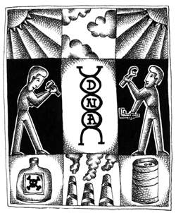
Are they good or are they bad? Genetic mutations, that is. The answer, like the answers to so many other questions in life is: It all depends. What it depends on is context, and that context has to do with Darwin and the ways in which cancer inhabits and inhibits the lives of our planet's beings.
 
It all starts with our day-to-day existence, when the DNA in living cells is exposed to a plethora of environmental effects ­ solar rays, chemical materials, the by-products of the metabolic process within the cells themselves, etc. Simply stated: Our DNA becomes all stressed out.

These stressful influences may consequently damage DNA, and are expressed in change, breakdown, and replacement of the genetic "letters" which comprise our genetically-encoded information. (DNA is built from millions of these genetic letters.) Damaged data leads to the creation of defective proteins, which may in turn alter the correct operation of the cell. Under certain circumstances, this can even lead to cell death.

Here's the lowdown on understanding how mutation works.
 
"Bad" mutations may activate cancer-causing genes, or "switch off" cancer-resistant genes; without the protection of resistance, there's a possibility of the development of cancerous growth. Other negative mutations are liable to jumpstart the processes which cause the appearance of other diseases.
 
On the other hand, "good" mutations form the steps upon which life ascends the evolutionary ladder. In fact, without mutation, evolution is impossible.
 
We're all familiar with that first evolutionary revolutionary, Darwin. While he didn't have access to the sophisticated research techniques of today, Darwin's work constituted a precursor to the field of genetics.
 
A century later, scientific research is delving into the wide genetic differences by which natural selection acts. Darwin certainly would have been intrigued to learn that selection is based on the accumulation of nonfatal mutations in hereditary DNA material within the nuclei of all living creatures. This process sometimes triggers an improvement in the survivability of living creatures.
 
That's where Prof. Zvi Livneh and his research students, Nina Reuven and Guy Tomer, of the Institute's Biological Chemistry Department, come into the evolutionary picture. With survivability the issue, the Weizmann Institute team is asking this question: How do you prevent and reduce, to whatever extent possible, the damage of the bad mutations on one hand, while on the other, preserve what is most beneficial about the evolutionary process?
 
What we do know is that the living, breathing world has its own built-in, sophisticated mechanisms that allow cells to repair defective DNA. However, these mechanisms aren't always perfect; they sometimes undertake their work with a certain degree of "negligence." This opens the possibility for other mutations, only some of which may be considered beneficial ones.
 
Mutations are incorrect coding instructions, formed when the replication machinery duplicates the DNA regions containing the unrepaired lesions. It was originally thought that replication stops when encountering damaged DNA, and that the process would continue only with the assistance of "helping proteins."
 
What Livneh and his team discovered is that the enzyme polymerase, which replicates the DNA, "didn't stop at the red light" when it came upon the damage ­ not even in the absence of the helping proteins.
 
So why are the helping proteins required at all?
 
First, the helpers make the replication of DNA damage go faster. But what Livneh and team found is yet another reason: The choice of the polymerase to replicate the damaged region either by itself, or with the assistance of the helping proteins, is a life or death decision.
 
Without the presence of helping proteins, the polymerase ignores DNA damage, leaving blank spaces of one or two letters in the DNA's genetic continuity. If DNA is like a book containing words made of letters that appear in triplet form, removing one or two of them sends a whole different message. For example, if you have the sentence "Dad did cry" and you remove "D" from "Dad," the message is read as "add idc ry." The message is clearly set askew.
 
This leads to the creation of shortened proteins that are liable to hasten the cell's death. When polymerase encounters damage and continues its action in the presence of helping proteins, it fills in the blanks created in the genetic continuity with random genetic letters. While this still leads to the creation of a mutation, it is a mild type and the mutant proteins can work, if only partially. In any event, they do not usually bring about the death of the cell. In fact, they may act like some of us do when we're under stress: We perform better. On occasion, the mutation can actually improve the protein.
 
Livneh: "We knew this before, but we didn't know the rationale for it. If the helping proteins aren't there, the replication machine is making disastrous mistakes."
 
Prof. Livneh's discovery: reconstituting the entire mutagenic process in a test tube by taking components from billions of living cells. Put them to work outside the cells.
Sit back a few minutes while the entire filling-in process happens in a matter of minutes.
 
What does Livneh's research have to do with life in the 21st century? Plenty. It's important to note that one way bacteria acquire resistance to antibiotics is due to mutation. Beneficially, one of the potential, long-range applications of Livneh's work is the possibility that some day, we may be able to counteract this scary news: We're running out of antibiotics that kill bacteria.
 
Under most circumstances, it is clear that mutations created as a reaction to conditions of stress, improve the ability of bacteria to survive these conditions. Prof. Livneh's research is likely to broaden the boundaries of our knowledge regarding the ways in which living creatures are able to adapt to environmental conditions through evolution, good or bad.
 
It all depends.
 

Many causes of DNA damage. illustration: Mutations

 
Life Sciences
English

Pages