<p>
Science Feature Articles</p>

A Case of Mistaken Identity

English


Prof. Mark Safro. Proofreading equipment


Considering that all of the 100,000 or so different proteins in our bodies are made of chains constructed of thousands of linked amino acids, the error rate is quite low. Yet now and again, the wrong bit of material will make its way onto the assembly line in the protein factory – the ribosome – and end up in the polypeptide chain. Since the sequence of amino acids determines the shape of the folded protein, as well as its function, even one wrong link in the chain can spell disaster for some proteins.

Prof. Mark Safro and his colleagues Drs. Nina Moor and Liron Klipcan of the Structural Biology Department in the Faculty of Chemistry recently revealed how one of the most common substitutions can slip right by the molecular equipment that is meant to prevent mistakes. Their findings may be relevant to many disorders, including Alzheimer’s disease.

Safro and his group investigate a central step in the complex protein manufacturing process – one that helps ensure the fidelity of the translation from instruction list to finished protein. This step involves an interaction between an adaptor RNA molecule called tRNA and an enzyme known as aminoacyl-tRNA synthetase. If the tRNA can be thought of as the “trucks” that carry the individual amino acids to the assembly line in the ribosome, aminoacyl-tRNA synthetase is the “loader” that puts the amino acids on the trucks. Each such loader is familiar with one amino acid, and this is the one it captures. But some synthetases have an added responsibility. Of the 20 amino acids from which all proteins are built, some of the building blocks are easily confused with one another, and the synthetases for these components have evolved an extra function, called “proofreading,” for double-checking the shipment before sending it on its way.


Levodopa in the active site of mitochondrial Phenylalanyl-tRNA synthetase. The lattice is the observed electron density corresponding to the L-Dopa
 
Such is the case with the amino acid tyrosine, which differs from a second amino acid, phenylalanine, only by the addition of two simple atoms – an OH group. To complicate matters, tyrosine can also be mistaken for a molecule that resembles an amino acid but has a totally different function. This molecule is levodopa, or L-Dopa, better known as the common treatment for Parkinson’s disease. L-Dopa resembles tyrosine because it is made in the body by adding yet another OH group to a tyrosine molecule. This small “quasi” amino acid is able to cross into the brain, where it is converted into dopamine – the neurotransmitter that is under-produced in Parkinson’s patients. But when L-Dopa is accidentally incorporated into a protein instead of tyrosine, that second OH group becomes a problem. This tiny addition is chemically active, and it can cause the proteins to clump together into aggregates that don’t break down easily.

Safro and his team realized that the question of L-Dopa inclusion was complicated by the fact that there are two types of aminoacyl-tRNA synthetase – one in the cell’s cytoplasm and one in organelles called mitochondria, the cell’s power plants. When the researchers crystallized both types to reveal their structures, they found that the mitochondrial synthetase is a bare-bones, stripped-down version of its counterpart in the cell body. Among other things, it lacks the proofreading equipment.

Next, the team asked how capable either version is of recognizing L-Dopa and preventing it from getting into the protein chain. Using a variety of experimental methods – including a tour de force of crystallization in which they succeeded in capturing the 3-D structures of synthetase and L-Dopa acting together, as well as kinetic experiments – they showed how the mistake occurs. It appears, says Safro, that in this particular instance, the proofreading mechanism is not up to the task. The L-Dopa assumes the same orientation in the synthetase as tyrosine and thus, to the proofreader, it can look identical. While failure to recognize the false amino acid was seen in both versions, it was especially apparent in the stripped-down mitochondrial synthetases – which, unfortunately, have a greater impact on human health.

Mistakes in protein assembly are fairly rare; L-Dopa appears to be a somewhat unique case of mistaken amino acid identity. But it may also be a critical one: Protein aggregates like the ones caused by faulty L-Dopa inclusion are implicated in Alzheimer’s disease, and Safro believes that this “blind spot” in the proofreading machinery may be an important contributing factor.
 
Prof. Mark Safro is the incumbent of the Lee and William Abramowitz Professorial Chair of Macromolecular Biophysics.
 
 
Levodopa in the active site of mitochondrial Phenylalanyl-tRNA synthetase. The lattice is the observed electron density corresponding to the L-Dopa
Space & Physics
English

Pumping Iron

English


(l-r) Dr. Ishai Dror and Prof. Brian Berkowitz. Clean water


Iron and vitamin B12 are no longer just for dietary supplements. When bound together with a third material, they can break down some of the worst water pollutants, and they may thus be the key to restoring the health of the underground water supply.

Using these components, Prof. Brian Berkowitz and Dr. Ishai Dror of the Environmental Sciences and Energy Research Department (Faculty of Chemistry) have designed a treatment system that reduces the toxic chemicals in water to harmless compounds that can be further broken down naturally. Such a system could be placed in the path of underground flow to clean the water before it ever reaches the pipes, or it could be used to treat polluted water pumped from below ground.

Many of the waste chemicals that leach down into underground water reservoirs, called aquifers, can persist there for hundreds, even thousands of years. The problem, says Berkowitz, is that such man-made substances as pesticides, cleaning fluids and flame retardants don’t resemble anything found in nature, and thus few natural mechanisms exist to break them down. Unfortunately, many are considered to be carcinogenic even in extremely minuscule amounts, and Western standards allow only a few parts per billion of these pollutants to be present in drinking water.

Berkowitz and Dror realized that a simple chemical reaction might split the molecules of these harmful substances apart in such a way that bacteria or other natural processes could finish the job. The “indigestible” part of these molecules is generally a bond between a carbon ring and a chloride or bromide ion; if the reaction could get them to accept an electron in place of the ion, the two would be separated. As an electron source, the researchers turned to elemental iron, which easily gives up its electrons. They then looked for a way to combine the iron with a common filtering material called diatomite. Diatomite, though it resembles very fine white, powdery sand, is actually composed of fossilized diatom shells, and the microscopic pores in the grains make it an excellent filter for fluids – everything from swimming pool water to beer.
 

Scanning electron microscope images of representative samples of (left) clean diatomite and (right) diatomite composite with zero valent iron (small white dots) and vitamin B12 (not visible)


At first, the iron didn’t bind well to the diatomite base; and the electron transfer was only partially successful. The scientists realized they needed a further addition to the mix – a catalyst that would help get the electrons from the iron into the polluting molecules. “Rather than invent a new catalyst, we looked to natural molecules,” says Dror. “Nature’s favorite molecule for shuttling electrons is the porphyrin.” Porphyrins are found in everything from chlorophyll to hemoglobin to vitamin B12. The scientists found that B12 – an easily available, non-toxic substance – was just what the doctor ordered. The vitamin kept the iron particles small and spaced evenly on the diatoms’ surface, creating a large surface area for the chemical reactions to take place while enabling the water to flow through the material. Now the electron transfer was highly efficient. The costs of such a system, says Berkowitz, are comparable to those of standard activated carbon filters, and the results are much better, making them attractive to industries that rely on clean water.  

Larger diatomite/iron/B12 units could be placed in trenches or batteries of deep wells dug into the path of water flowing underground, says Berkowitz. In this way, aquifers that are in danger of contamination could be restored and their precious water reserves saved. Added advantages might include the prevention of soil contamination through seepage, a reduction in the need for such energy-intensive water-purification methods as desalination and a halt to the release of toxic gases from polluted aquifers into underground basements and parking garages.
 
Prof. Brian Berkowitz’s research is supported by the Carolito Stiftung; the Angel Faivovich Foundation for Ecological Research; the Dr. Scholl Center for Water and Climate, which he heads; the Brita Fund for Scholarships, Research and Education for the Improvement of Water in Israel; the Phyllis and Joseph Gurwin Fund for Scientific Advancement; Mr. and Mrs. Michael Levine, Pinckney, NJ; and the P. & A.Guggenheim-Ascarelli Foundation. Prof. Berkowitz is the incumbent of the Sam Zuckerberg Professorial Chair in Hydrology.


Scanning electron microscope images of representative samples of (left) clean diatomite and (right) diatomite composite with zero valent iron (small white dots) and vitamin B12 (not visible)
Environment
English

Eat, Prey, Rain

English

Dr. Ilan Koren. In the clouds. Cloud photo: Tamar Deutsch


What do a herd of gazelles and a fluffy mass of clouds have in common? A mathematical formula that describes the population dynamics of prey animals, such as gazelles, and their predators has been used to model the relationship between cloud systems, rain and tiny floating particles called aerosols. This model may help climate scientists understand, among other things, how human-produced aerosols affect rainfall patterns.

Clouds are major contributors to the climate system. In particular, the shallow marine stratocumulus clouds that form huge cloud decks over the subtropical oceans cool the atmosphere by reflecting part of the incoming solar energy back to space. Drs. Ilan Koren of the Weizmann Institute’s Environmental Sciences and Energy Research Department (Faculty of Chemistry) and Graham Feingold of the NOAA Earth System Research Laboratory, Colorado, found that equations for modeling prey-predator cycles in the animal world were a handy analogy for cloud-rain cycles: Just as respective predator and prey populations expand and contract at each other’s expense, so too does rain deplete clouds, which grow again once the rain runs out. And just as the availability of grass affects herd size, the relative abundance of aerosols – which “feed” the clouds as droplets condense around them – affects the shape of those clouds. A larger supply of airborne particles gives rise to more droplets, but these droplets are smaller and thus remain high up in the cloud rather than falling as rain.

In previous research, Feingold and Koren had “zoomed in” to discover oscillations in convective cells in marine stratocumulus systems. Now they returned to their data, but from a “top-down” angle to see if a generalized formula could reveal something about these systems. Using just three simple equations, they developed a model showing that cloud-rain dynamics mimic three known predator-prey modes. Like gazelles and lions, the two can oscillate in tandem, the “predator” rain cycle following a step behind peak cloud formation. Or the two can reach a sort of steady state in which the clouds are replenished at the same rate as they are diminished (as in a light, steady drizzle). The third option is chaos – the crash that occurs when predator populations get out of hand or a strong rain destroys the cloud system.

The model shows that as the amount of aerosols changes, the system can shift abruptly from one state to another. The model shows a bifurcation –  two scenarios at different ends of the aerosol scale that lend themselves to stable patterns. In the first, relatively low aerosol levels lead to clouds in which development depends heavily on aerosol concentrations. In the second, high aerosol levels produce saturation; these clouds depend solely on the initial environmental conditions.

Using this so-called systems approach, says Koren, “can open new windows to view and understand the emergent behavior of these systems and the complex relationships between clouds, rain and aerosols, giving us a useful view of the big picture and helping us to understand how shifting aerosol levels can lead to different climate patterns.”
 
Dr. Ilan Koren’s research is supported by the Yeda-Sela Center for Basic Research. Dr. Koren is the incumbent of the Benjamin H. Swig and Jack D. Weiler Career Development Chair in Perpetuity.
 
 
Dr. Ilan Koren. In the clouds. Cloud photo: Tamar Deutsch
Environment
English

Built to Scale

English


The patterns on these butterfly wings are perfectly scaled, even though they are different sizes


The wing of a fly is a marvelous piece of work; its unique properties rely, in part, on an intricate network of structural veins. If we compare a small fly wing to a large one, exactly the same pattern of veins – scaled to fit – appears. How does this happen? For that matter, how do humans and animals grow limbs just the right size for their bodies? How do developing embryos keep everything in proportion as they grow?
 
Prof. Naama Barkai
 
These questions are ones of scaling – how patterns stay in sync with size as an embryo or organism grows and develops. Institute scientists Profs. Naama Barkai and Ben-Zion Shilo and research student Danny Ben-Zvi of the Molecular Genetics Department have now shown how scaling works in fruit fly wings, and their findings should be applicable to many different examples of development.

These researchers drew their inspiration from a famous experiment performed in the 1920s by German scientist Hans Spemann, in which he grafted cells from the future head of one frog embryo onto the flank of a second embryo. The resulting tadpole was a “Siamese twin” with two perfectly formed – but half-sized – heads. This finding implied that, rather than proceeding according to a pre-set “plan,” development takes place through cells sending messages to one another, directing them down certain developmental paths. In the intervening years, it has been found that the control mechanism for patterning relies on one such message – a substance called a morphogen. Morphogens are secreted by a small number of cells in the center of the developing embryo, and from there, they diffuse outward. As this substance disperses, its levels drop off in the cells farther from the center, and thus the morphogen concentration relays a signal to the developing cell about its place and function in the growing organism.


Prof. Ben-Zion Shilo. Above: Prof. Naama Barkai
 
But the Institute researchers knew this was not the whole story: A morphogen diffuses from the center at the same rate in a small organism as in a larger one and thus would not effect scaling on its own. Several years ago, they recreated Spemann’s frog embryo experiment, but they looked for a second signal – one that could bring size into the picture. Indeed, they found a molecule that is synthesized at the edges – one that also diffuses away from its site of origin. The second molecule functions as a morphogen, and it is the redistribution of this molecule that finally determines the morphogen signal each developing cell receives, in a way that also takes embryo size into consideration.

Next, they asked: Do all complex organisms use a similar scaling method? To explore this question, Barkai and Ben-Zvi first created a theoretical model in which a molecule on the growing edge – called an expander molecule – aids in the distribution of the morphogen. The morphogen is referred to in the model as a repression molecule because it also shuts down the synthesis of the expander molecule at the edge. The model suggests just how this interplay between expansion created at the edge and repression moving from the center results in a pattern built to scale.
 
In the new study, which appeared in Current Biology, Ben-Zvi, Barkai and Shilo have brought the theoretical model back into the lab, carrying out experiments on the development of wings in fruit fly larvae. These larvae don’t actually have wings; nevertheless, wing patterning is already taking place in small structures called wing discs, and their development can easily be traced. The researchers knew that a morphogen called Dpp was active in the wing disks, and they suspected that a protein called “Pentagone,” acted as an expander molecule, as it had recently been shown to play a role in the development of wing vein networks.

The scientists collected fruit fly larvae of varying size and, using a quantitative method they developed, checked the distribution of morphogen concentrations. They then eliminated Pentagone from the developing wing and checked again. Their findings showed that the wings in the unaltered fruit flies revealed the morphogen scaling activity predicted in the model, its signals being proportionate to wing size. In contrast, in the flies without Pentagone the morphogen was distributed in the same way in all the wings, regardless of their relative sizes. They were thus able to show that Pentagone is, indeed, an expansion molecule and that the expansion-repression paradigm they had formulated can be applied to such differing organisms as frogs and fruit flies.

Shilo: “Frog embryos and fly wings in larvae develop through completely different molecular mechanisms, yet they apparently follow the same general scaling model. The beauty of this research lies in the way it seamlessly weaves a theoretical model into experimental biology. This gives us a fresh approach to investigating scaling, an approach in which, rather than getting bogged down in a search for complex molecular mechanisms, we can begin by looking for this relatively simple and universal paradigm.”
 
Prof. Naama Barkai’s research is supported by the Helen and Martin Kimmel Award for Innovative Investigation; the Jeanne and Joseph Nissim Foundation for Life Sciences Research; the Carolito Stiftung; Lorna Greenberg Scherzer, Canada; the Minna James Heineman Stiftung; and the European Research Council. Prof. Barkai is the incumbent of the Lorna Greenberg Scherzer Professorial Chair.

Prof. Ben-Zion Shilo’s research is supported by the Jeanne and Joseph Nissim Foundation for Life Sciences Research; the Dr. Josef Cohn Minerva Center for Biomembrane Research; the Carolito Stiftung; la Fondation Raphael et Regina Levy; the estate of Georg Galai; and the Mary Ralph Designated Philanthropic Fund. Prof. Shilo is the incumbent of the Hilda and Cecil Lewis Professorial Chair of Molecular Genetics.


The patterns on these butterfly wings are perfectly scaled, even though they are different sizes
Life Sciences
English

Identifying Autism Early

English
 
Prof. Rafael Malach and Dr. Ilan Dinstein. Synchronization


The biological causes of autism are still not understood. A diagnosis of autism is possible only after age three or four; and the tests are subjective, based on behavioral symptoms. Now, in research that appeared in Neuron, scientists at the Weizmann Institute of Science, Carnegie Mellon University and the University of California, San Diego, have found, for the first time, a method that can accurately identify a biological sign of autism in very young toddlers. By scanning the brain activity of sleeping children, the scientists discovered that the autistic brains exhibited significantly weaker synchronization between brain areas tied to language and communication, compared to that of non-autistic children.
 
As compared to the control brain (top), the autistic brain (bottom) shows weaker inter-hemispheric synchronization in several areas, particularly the superior temporal gyrus (light blue) and the inferior frontal gyrus (red)
 
“Identifying biological signs of autism has been a major goal for many scientists around the world, both because such signs may allow early diagnosis and because they can provide researchers with important clues about the causes and development of the disorder,” says postdoctoral fellow Dr. Ilan Dinstein, a member of the group of Prof. Rafael Malach, who headed this study in the Weizmann Institute’s Neurobiology Department.
 
While many scientists believed that faulty lines of communication between different parts of the brain were involved in the spectrum of autism disorders, there was no way to observe this in very young children, who are unable to lie still inside an fMRI scanner while they are awake. But work by Malach’s group and other research groups pointed to a solution. Their studies had shown that even during sleep, the brain does not actually switch off. Rather, the electrical activity of the brain cells switches over to spontaneous fluctuation. These fluctuations are coordinated across the two hemispheres of the brain such that each point on the left is synchronized with its corresponding point in the right hemisphere.

In the sleeping autistic toddlers, the fMRI scans showed lowered levels of synchronization between the left and right brain areas known to be involved in language and communication. This pattern was not seen either in children with normal development or in those with delayed language development who were not autistic. In fact, the researchers found that this synchronization was strongly tied to the autistic child’s ability to communicate: The weaker the synchronization, the more severe were the symptoms of autism. On the basis of the scans, the scientists were able to identify 70% of the autistic children between the ages of one and three.

Dinstein: “This biological measurement could help diagnose autism at a very early stage. The goal for the near future is to find additional markers that can improve the accuracy and the reliability of the diagnosis.”
 
Prof. Rafael Malach’s research is supported by the Nella and Leon Benoziyo Center for Neurosciences, which he heads; the Nella and Leon Benoziyo Center for Neurological Diseases; the Carl and Micaela Einhorn-Dominic Brain Research Institute; the Kahn Family Research Center for Systems Biology of the Human Cell; the Friends of Dr. Lou Siminovitch; and the S. & J. Lurje Memorial Foundation. Prof. Malach is the recipient of the Helen and Martin Kimmel Award for Innovative Investigation and he is the incumbent of the Barbara and Morris L. Levinson Professorial Chair in Brain Research.
 
 
As compared to the control brain (top), the autistic brain (bottom) shows weaker inter-hemispheric synchronization in several areas, particularly the superior temporal gyrus (light blue) and the inferior frontal gyrus (red)
Life Sciences
English

Excitement That Shows

English


laser


Chemistry is a messy business. Complex chemical reactions typically involve numerous steps and often generate toxic waste. Transforming one chemical substance into another usually demands mixing different materials, supplying energy, adding catalysts. But chemists have a dream: One day, they hope to run chemical reactions simply by directing a laser beam at a target molecule, selectively breaking a few chemical bonds to obtain the desired product.

To the dismay of alchemists and others, this method won’t turn lead into gold (rather than breaking chemical bonds, such a change would require altering the atomic nucleus, which cannot be accomplished with lasers), but it can have a host of other advantages: Laser control can render chemical reactions effective, clean and cheap. For example, certain drugs currently produced by complex organic chemistry processes requiring several dozen transformations might in the future be obtained by considerably simpler and quicker methods.

Prof. David Tannor of the Weizmann Institute’s Chemical Physics Department, who has studied laser control of chemical reactions for more than a quarter of a century, believes the method may lead to future commercial applications. His optimism stems from the fast pace of research in leading labs around the world, including his own. Recently, Tannor and graduate student David Avisar accomplished a major feat: They developed a systematic theoretical method for reconstructing the geometrical changes occurring in a molecule after it shifts into an “excited,” high-energy state as a result of exposure to radiation. Molecules in this state emit light at characteristic times and wavelengths. As reported in Physical Review Letters, the scientists have analyzed this light in order to trace, in retrospect, what happened in the molecule during excitation. “What we’ve done is equivalent to reconstructing the cow by looking at the hamburger,” says Tannor.
 
 
Prof. David Tannor. Laser control
 
When molecules become excited, they undergo a variety of changes: They might be stretched or twisted, and some of their chemical bonds might be broken. The Weizmann Institute reconstruction, performed by a sequence of mathematical transformations, reveals the exact sequence of events that take place in a two-atom molecule of lithium from the time it gets excited until it falls apart. The scientists are now planning to perform similar reconstructions with larger molecules made up of three or more atoms.

Such reconstruction can be a boon to laser-control chemistry because it provides crucial information about the changes induced in molecular structure by light, which in turn can make it possible to manipulate chemical bonds on demand. If the scientists know which bonds are weakened or broken following the excitation by light, they can make the process more efficient and selective with the help of specially designed laser pulses. In addition, the new understanding of excited molecules can advance the study of matter in the atmosphere and stratosphere, where a wide variety of molecules exposed to solar radiation exist in an excited state.


Beware of Traps


One major stumbling block to achieving laser control of chemical reactions is efficiency. If, say, the control is only 50 % efficient – that is, if only half of the product obtained in the reaction is the desired substance – the method will be of limited use. The goal is to reach an optimal efficiency, as close as possible to 100%.

Until recently, scientists believed that, theoretically, these high efficiencies were virtually guaranteed, provided one was persistent enough: Keep improving the precision of laser control and ultimately you will reach the optimal level. But in a study recently published in Physical Review Letters, Prof. David Tannor and research associate Dr. Alexander N. Pechen have shown this assumption to be false. It turns out that in certain cases, the improvement hits a dead end: What looks like a steady increase for a while, ultimately peaks at a level that falls far below 100 %. In such cases, the initial progress leads to a “trap”: Since no further improvement is possible, the scientist has no choice but to abandon the entire process and begin all over again from a different starting point.

The bad news is that these newly discovered traps make optimal efficiency more difficult to reach. The good news is that knowing about them can help scientists avoid them – for example, by applying strict criteria to the starting parameters in the improvement process.
 
Prof. David Tannor is the incumbent of the Hermann Mayer Professorial Chair.
 
Chemistry
English

View from the Top

English

Drs. Anna and Dmitry Gourevitch. Love and math


Even as a child, Dr. Dmitry Gourevitch knew he was going to study the exact sciences. But when he embarked on university studies at age 15, after immigrating to Israel from Russia, he wasn’t sure he was going to opt for an academic career. All doubts disappeared when he encountered representation theory, a highly abstract area of mathematics. “It felt as if I’d climbed a mountain peak and could observe parts of the Earth I’d never seen before,” recalls Gourevitch, who recently joined the Weizmann Institute’s Mathematics Department. “The joy of discovery was tremendous; I suddenly realized how various concepts were interconnected in ways I had no idea existed.”

Gourevitch has been seriously engaged in mathematics for more than half of his 29 years, thanks in large part to the school he attended in St. Petersburg (then known as Leningrad). The legendary school No. 30 was known to be a breeding ground for mathematical geniuses: Its students won a disproportionate number of prizes at local and national mathematics Olympiads, many of them later becoming professional mathematicians.

After “making aliya” with his parents in 1995, Gourevitch entered a “special status” program allowing him to study towards a degree in mathematics at Tel Aviv University in parallel with high school. It was in a university course taught by Prof. Joseph Bernstein that he was struck by the explanatory power of algebra, particularly representation theory. He was ultimately to make this theory the focus of his research – but only after earning his B.Sc. with honors at age 18, completing his compulsory service in the Israel Defense Forces and, at 22, enrolling in graduate studies at the Weizmann Institute. He earned his M.Sc. and Ph.D. degrees from the Institute under the guidance of Bernstein and Weizmann’s Prof. Stephen Gelbart. After conducting postdoctoral research at the Institute for Advanced Study in Princeton and at Rutgers, New Jersey, Gourevitch now works on representation theory in his position of Senior Scientist at Weizmann.

Representation theory, which studies the symmetries of linear spaces, is so powerful because it combines two of the most fundamental concepts in mathematics: the technique of linearization and the notion of symmetry, which appears in multiple problems in mathematics, physics and other exact sciences. The theory has a wide variety of applications – in other areas in mathematics, in quantum mechanics, in engineering and in computer science. Translating basic research into practice, however, takes time. Many decades passed, for instance, before certain concepts of representation theory, developed in the late nineteenth and early twentieth centuries, found their application in computer tomography (CT) – the medical imaging technique. Similarly, it took decades before other concepts helped to speed computation or provide solutions to major problems in computer science, such as the expander problem, whose solution allows one to connect multiple users into a single communications network in an optimal manner.

It’s impossible to predict what applications might one day arise from Gourevitch’s current research. Together with colleagues, he has already managed to prove the so-called multiplicity one conjectures, which had remained unproven for 20 years. He has also made several other contributions to his field and is hoping to make more. The one thing that’s certain is that the mountain peak he is climbing in his studies is particularly high – he is working on representations of the so-called non-compact groups, notorious for their complexity – and that it is bound to help reveal new landscapes that are presently hidden from the scientists’ view.

Mathematical romance

Dmitry and his wife Anna met at a lecture in algebraic topology at Tel Aviv University. Their romance was fostered by a shared interest in algebraic geometry. Shortly after getting married, the couple published a joint paper in the Journal of Pure and Applied Algebra. They now live in Rehovot with their children. Anna is a teaching associate in mathematics at Tel Aviv University, from which she earned her Ph.D.
 
 
Drs. Anna and Dmitry Gourevitch. Love and math
Math & Computer Science
English

Fast Focus

English
 
 
(l-r) Ori Katz, Eran Small and Prof. Yaron Silberberg. Under the skin


A few well-known facts about lasers: These ultra-focused beams of light have been used for decades to cut metal cleanly and precisely. In the field of medicine they take the place of sharp surgical scalpels; they are also used in some kinds of medical imaging; and they are a part of today’s advanced optical microscopes. Many of these uses require the light to be focused tightly to a very narrow, highly intense point. One way of cutting cleanly without causing harm to the surrounding area – say, in biological tissue that is easily damaged by excess energy – is to zap the target point with very brief (less than a millionth of a millionth of a second) flashes of highly concentrated laser light.


This is all well and fine when the target point is on an exposed surface. But scientists and surgeons would like to be able to aim lasers under the skin as well – for instance at tumors inside the body. And herein lies the difficulty: A standard laser can’t focus even a millimeter under the skin. For one thing, biological tissue is simply not transparent – meaning the light entering it scatters in all directions. When it passes through tissue, a flash of laser beam that is extremely short and focused when it leaves its source tends to lengthen and spread out along the way. Its intensity weakened, the beam loses its scalpel-like ability to slice cleanly through living matter.
 
 
Fluorescence created by a flash of laser light passing through a ½ mm piece of bone before (l) and after (r) application of the algorithm to focus the beam to a single point
 
Prof. Yaron Silberberg and research students Ori Katz, Eran Small and Yaron Bromberg of the Physics of Complex Systems Department sought a way to focus rapid flashes of laser light as they pass through a scattering layer. The method they developed works on feedback: They created a system that can assess, in real time, how the light scatters. Using algorithms they developed, they created a beam that can “anticipate” the dispersal of its light and make the necessary corrections. This computerized system is able to tailor a laser beam to the tissue such that it is precisely and narrowly focused on the internal target.  

In this research, which appeared in Nature Photonics, the scientists employed a simple LCD screen, similar to those found in computer projectors, to correct the beams’ focus. While it was known that screens of this type could be used to correct spatial errors, Silberberg and his team succeeded in demonstrating that this simple system can also correct for errors in the dimension of time.

The scientists hope that this system will, in the future, aid in the development of applications, including new types of medical lasers and optical microscopes that will enable researchers in the life sciences to get under the skin and into the underlying tissue.
 
Prof. Yaron Silberberg’s research is supported by the Crown Photonics Center, which he heads; the Wolfson Family Charitable Trust; and the Cymerman - Jakubskind Prize. Prof. Silberberg is the incumbent of the Harry Weinrebe Professorial Chair of Laser Physics.
 
 
(l-r) Ori Katz, Eran Small and Prof. Yaron Silberberg. Under the skin
Space & Physics
English

Thanks for the Memories

English

fMRI


How easy is it to falsify memories? New research at the Weizmann Institute shows that a bit of social pressure may be all that is needed. The study, which appeared recently in Science, reveals a unique pattern of brain activity when false memories are formed – one that hints at a surprising connection between our social selves and memory.

The experiment, conducted by Prof. Yadin Dudai and research student Micah Edelson of the Institute’s Neurobiology Department, together with Prof. Raymond Dolan and Dr. Tali Sharot of University College London, took place in four stages. In the first, volunteers watched a documentary film in small groups. Three days later, they returned to the lab individually to take a memory test, answering questions about the film. They were also asked how confident they were about their answers.

They were later invited back to the lab to retake the test while being scanned in a functional magnetic resonance imager (fMRI) that revealed their brain activity. This time, the subjects were also given a “lifeline”: the supposed answers of the others in their film-viewing group (along with social-media-style photos). Planted among these were false answers to questions the volunteers had previously answered correctly and confidently. After seeing these “planted” responses, the participants conformed to the group, giving incorrect answers nearly 70% of the time.  

But were they simply conforming to perceived social demands, or had their memory of the film actually undergone a change? To find out, the researchers invited the subjects back to the lab to take the memory test once again, telling them that the answers they had previously been fed were not those of their fellow film watchers but random computer generations. In some cases the responses reverted back to the original, correct ones; but close to half remained erroneous, implying that the subjects were relying on false memories implanted in the earlier session.

An analysis of the fMRI data showed differences in brain activity between the persistent false memories and the temporary errors of social compliance. The most outstanding feature of the false memories was a strong co-activation and connectivity between two brain areas: the hippocampus and the amygdala. The hippocampus is known to play a role in long-term memory formation, while the amygdala, sometimes known as the emotion center of the brain, plays a role in social interaction. The scientists think that the amygdala may act as a gateway connecting the social and the memory processing parts of the brain; its “stamp” may be needed for some types of memories, to give them approval before they get uploaded to the memory bank. Thus social reinforcement could act on the amygdala, persuading our brains to replace a strong memory with a false one.


Prof. Yadin Dudai’s research is supported by the Norman and Helen Asher Center for Human Brain Imaging, which he heads; the Nella and Leon Benoziyo Center for Neurological Diseases; the Carl and Micaela Einhorn-Dominic Institute of Brain Research, which he heads; the Marc Besen and the Pratt Foundation, Australia; Lisa Mierins Smith, Canada; the Abe and Kathryn Selsky Memorial Research Project; and Miel de Botton, UK. Prof. Dudai is the incumbent of the Sara and Michael Sela Professorial Chair of Neurobiology.
 
 
fMRI
Life Sciences
English

Supernova News

English


Before and after images of the Messier 100 Galaxy reveal the appearance of SN 2006X, one of the supernovae used in the study. Photos: European Southern Observatory (ESO)


Cosmic distances are difficult to grasp and no less difficult to measure. When it comes to other galaxies or even remote parts of our own Milky Way, distance measurements are nothing but assessments, derived from indirect clues.
 
Highly important among such clues are supernovae, extremely luminous stellar explosions. The distance to a supernova of a particular type, called Type Ia, can be calculated from its brightness: the brighter it appears, the closer it is to the viewer. Thanks to such supernovae, for example, astronomers have famously revealed that our universe is expanding at an accelerated pace, which suggests that it’s permeated with mysterious dark energy. These calculations, however, are based on the assumption that all Type Ia supernovae have the same luminosity. Are all these explosions indeed created equal?
 
Type Ia supernovae are thought to be born when an exceedingly dense star called a white dwarf receives more mass from a nearby star, until it’s so “overwhelmed” that it explodes. A new study reported in Science and led by Weizmann Institute researchers, has gained major insight into the nature of these mass “donors.” The study was performed by Dr. Avishay Gal-Yam and postdoctoral fellow Dr. Assaf Sternberg of Weizmann’s Particle Physics and Astrophysics Department, in collaboration with scientists from more than a dozen research centers in the United States, Europe and Australia.
 
The researchers have revealed that in about a quarter of the cases in spiral galaxies (like the one pictured above), and possibly more, the companion star that “donates” its mass to the white dwarf is probably a regular, medium-sized star, largely similar to our own Sun. They reached this conclusion by analyzing the outflow of gas, typical of sun-like stars, observed during the “donation” of the mass. These findings constitute a major step toward determining the nature of all stellar “donors,” with the ultimate goal of establishing whether supernovae everywhere evolve in the same manner, having the same luminosity at various stages. Understanding their evolution, in turn, can greatly enhance our ability to measure distances throughout the cosmos and map its evolution and geometry.
 
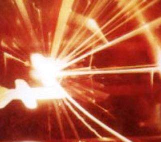
Institute observatory captures images of a new supernova
 
Photo: Ilan Manulis, Martin Kraar Observatory
 

Exploding stars are the "factories" that produce all the heavy elements found, among other places, in our bodies. In this sense, we are all stardust. These exploding stars – supernovae – are highly energetic events that can occasionally light up the night sky. Such an explosion generally disrupts the balance between gravity – which pulls the star's material inward – and the thermonuclear reaction at the star's core – which heats it and pushes it outward.

Certain types of stars to which this happens have 10-100 time the mass and are much younger than our sun. In them, the nuclear reaction begins like that of our sun – fusing hydrogen into helium – but the fusion then continues, producing heavier and heavier elements. The nuclear reaction eventually stops with iron, as there is no energy benefit to the star to fuse the heavier atoms, and the balance between gravity and thermonuclear activity comes to a halt. Gravity then takes over, and the mass of the star collapses quickly, releasing so much energy in the process that the explosion ensues. The star hurls its outer layers into space, and a new "bright star" appears in the night sky where none was seen before. Just such a new star was observed in the night sky between May 31 and June 1 in a spiral arm of our galaxy's close neighbor, the M51 galaxy.

The first to identify the supernova were amateur astronomers in France, and soon after it was detected by the Palomar Transient Factory (PTF) Sky Survey, in which Weizmann Institute scientists participate (see below). The phenomenon was also photographed in the new Martin Kraar Observatory at the Weizmann Institute, as well as in Tel Aviv University's Wise Observatory in Mitzpe Ramon. Israel's place on the globe enables its scientists to follow supernova events when it is daytime for many other observers, and thus to add significantly to the data collection.

The new supernova is being studied by an international team of researchers, including Dr. Avishay Gal-Yam and his research team, Drs. Ofer Yaron, David Polishook and Dong Xu, research students Iair Arcavi and Sagi Ben Ami, and Director of the Kraar Observatory, Ilan Manulis, all of the Weizmann Institute's Particle Physics and Astrophysics Department, as well as scientists from the US, England, Canada and other countries. They have already noted that the material thrown into space in the explosion contains a wide variety of elements. The mix they observed is atypical of supernova events at such an early stage of the explosion, and they plan to investigate this phenomenon.   


The last supernova observed in M51 (which is a mere 26 million light years away) occurred in 2005. Supernovae are thought to appear about once in 100 years in any given galaxy. The high occurrence in M51 can be explained by its interaction with a nearby galaxy, which causes the process of massive star formation to accelerate, thus increasing the rate of collapse and explosion as well.


Brighter, hotter, faster


A new class of supernova has scientists baffled. Recently, astrophysicists participating in the Palomar Transient Factory (PTF) – a project based at the Palomar Observatory in California that searches for transient flashes of light indicating stellar explosions – identified four new supernovae that did not conform to any known patterns. The PTF is an international collaboration in which the Weizmann Institute is a founding partner. The Institute’s Dr. Avishay Gal-Yam and research student Iair Arcavi, and Dr. Eran Ofek of the California Institute of Technology, who will soon be joining the Weizmann Institute, were part of the team that identified what makes these exploding stars so unique. In the process, they realized that another two, discovered earlier, also fit the pattern, bringing the number of supernovae in the new class to six.

So far, they have found that these supernovae have very little hydrogen – a puzzle, since most stars throw out large amounts of hydrogen when they explode. In addition, they are about ten times brighter than the most common type of supernova, and hotter as well; they expand at a rate of about 10,000 kilometers a second; and they take much longer to fade away than the others.

The processes that shape these new supernovae can’t be explained by the existing models, though several explanations have been proposed. Strangely, all were found in dwarf galaxies, and the explosions lit up these dim galaxies, illuminating the hard-to-see star groups and possibly shedding light on the fate of ancient, massive stars in the early Universe.
 

(l-r) Ilan Manulis, Dr. Avishay Gal-Yam, Dr. Ofer Yaron and Iair Arcavi. Explosive new images

Dr. Avishai Gal-Yam’s research is supported by the Nella and Leon Benoziyo Center for Astrophysics; the Yeda-Sela Center for Basic Research; the Peter and Patricia Gruber Award; the Legacy Heritage Fund Program of the Israel Science Foundation; Miel de Botton Aynsley, UK; and the Lord Sieff of Brimpton Memorial Fund.

Before and after images of the Messier 100 Galaxy reveal the appearance of SN 2006X, one of the supernovae used in the study. Photos: European Southern Observatory (ESO)
Space & Physics
English

Pages