Are you a journalist? Please sign up here for our press releases
Subscribe to our monthly newsletter:
Weizmann Institute scientists present a new method for imaging individual electrons

A new method for mapping gene expression deep in the body could one day be used to track stem cells or cancer therapy
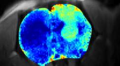
The method revealed tissue disruptions that cannot be seen by other means

A “crazy idea” for using fluoride nanocrystals as magnetic resonance tracers could lead to new clinical imaging methods
 containing the optic nerve indicated by the yellow arrows")
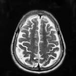
A new MRI method developed at the Weizmann Institute of Science improves our ability to study the brain and other non-homogeneous tissues
; the left hemisphere remained intact (green square)")
An MRI method for picking up the faintest signals can reveal the workings of the brain

A new MRI technique reveals how the mother's blood flow and that of the fetus meet in the placenta

A new MRI-based method can detect metabolite levels in real time
 Brain proton MRI; (c) mean cellular size; (r) distribution peak")
From quantum physics to biology, a new approach to magnetic resonance turns protons into “spies”