- Benefits of the big chill
- Moving on up
- When blood cells die
- Muscle building
- Preparation for the beginning of life
- The egg's secret attraction
- To bind or not to bind?
Benefits of the big chill
The electron microscope is the main research tool allowing examination of minute structures in cells and their surroundings. Unfortunately, standard electron microscopes operate in a vacuum and are only suited to examining dry preparations, which involve dehydrating cells and possibly damaging the structural integrity of the specimens examined.
In order to overcome this difficulty, Weizmann Institute scientists developed a new method of electron microscopy that can use frozen tissue specimens. The new method is based on two innovations: firstly, freezing tissue specimens and preparing very thin sections; secondly, transferring the frozen specimen under the microscope and examining it at low temperatures, observing it from different angles. This method, in combination with computerized structural reconstructions, allows researchers to study the spatial structure of the different components of the cell and to investigate the location and function of the various intracellular systems.
Weizmann Institute scientists, in collaboration with colleagues at the University of California at San Francisco, developed a system of multidimensional microscopy allowing the collection and processing of information on dynamic processes in living cells and in complex organisms. The method is based on a digital imaging system for living cells and embryos that acquires pictures of all depths as a function of time, probing simultaneously several molecular components that are labeled with different fluorescent dyes. This computerized system processes the images and examines the changes taking place over the course of time. A multidimensional movie reflecting cellular events allows scientists to study cell function in real time.
A Weizmann Institute scientist discovered a protein, called vinculin, that is located on the inner aspect of the cell membrane precisely where the cytoskeleton, the cell's structural protein network, is attached. At these spots, the cell also binds to adjacent cells or to the extracellular matrix on which it grows. This creates a sort of mechanical bond and continuity between the cytoskeletons of many cells, allowing dispersal of the forces emanating, for example, from external pressure on the tissue or from internal forces produced by the cells.
Vinculin apparently also fulfills an important role in intercellular communication. Weizmann scientists discovered that when cells contact an external surface, a chain of reactions is locally triggered whereby specific enzymes add phosphate groups (phosphorylates) to each other and to additional proteins located in their vicinity. This chain transfers signals from the surface of the cell to the nucleus, activating specific cellular communication processes, such as those which trigger cells to divide or differentiate. This research is vital to understanding how living cells operate.
Weizmann Institute scientists became the first to visualize, in real time, a communication pathway that transmits messages from the cell surface to its genetic material (DNA) in the nucleus. These external messages influence the genes' expression in the cell and because of this, the cell's output, development, and behavior.
The communication pathway from the cell's surface to the nucleus is based on a chain reaction, in which one molecule phosphorylates a second, and that in turn phosphorylates a third and so on, until the last one in the chain transmits the message to the DNA (phosphorylation cascade). This can result, for example, in the expression of a specific gene which then induces production of protein that may alter the cell's structure and function, possibly as part of the development of an organ or other part of the whole organism.
Weizmann Institute scientists developed unique antibodies which identify and bind solely to a phosphorylated version of a central molecule of the communication pathway. This allowed the researchers to identify when and where a communication message is transmitted along the pathway, and to monitor how different messages (hormones) reaching the cell from outside influence the cell's behavior and development. The communication pathway studied is involved in the control of cell differentiation and proliferation. When it goes awry, it may cause cancerous growths.
Moving on up
The smallest locomotive motors in nature are situated in the coat of bacteria (of the Escherichia coli type). They move the flagella, the "hairs" that allow bacteria to move around. Bacteria can change direction by rotating the flagella clockwise or counterclockwise, thereby approaching a source of food or avoiding noxious surroundings. The motor that drives the flagellum changes its direction of rotation according to the state of a "switch" composed of three proteins that operate a minute gear box. Weizmann Institute scientists discovered the mode of operation of a protein which, when bound to the switch, shifts the direction of rotation of the flagellar motor.
When blood cells die
Weizmann Institute scientists used red blood cells as a model to research aging processes. Using an electron microscope, the scientists discovered and characterized the structural differences between the membranes of young and old red blood cells and found that the major change occurs near the end of their life span and that only very few cells in a regular blood sample have undergone this change.
The scientists then revealed that in diseases in which red blood cells die early, there are more blood cells whose membranes have the aged structure. A similar phenomenon was observed in the blood of patients suffering from a shortage of a specific enzyme, resulting in their sensitivity to foods and medications known to cause the destruction of red blood cells.
Additional research showed that the structural change in the red blood cell membranes causes a loss of part of the electrical charge on their surface and that this phenomenon initiates a chain of events that results in the old and injured cells being removed from the blood system.
Muscle building
Weizmann Institute scientists have developed innovative methods to grow muscle cells in tissue cultures outside of the body (in vitro). Thanks to these methods, Weizmann Institute scientists and their colleagues in other laboratories worldwide succeeded in elucidating the cellular and molecular events involved in muscle differentiation, including the isolation of genes which encode proteins important in muscle formation. These methods later helped in studying the control processes of genes expressed in the differentiation of muscle cells, as well as various muscle diseases, including muscular dystrophy.
Preparation for the beginning of life
Weizmann Institute scientists pioneered a method to maintain and research mammalian ovarian follicles, which include the developing egg, outside the body. This opened the way for direct identification of the hormones required for ovulation, including maturation of the egg and its preparation for fertilization, embryonic development, change of the follicle to the corpus luteum (the "yellow body" that secretes progesterone, an essential hormone for pregnancy), culminating in the release of the mature ovum from the follicle.
In later studies, Institute scientists identified the mechanisms that regulate the activity of protein-denaturing enzymes, allowing release of the ovum from the follicle and its transfer to the fertilization region. In these studies, the great similarity between the ovulation and infection processes was uncovered and it was demonstrated that interleukin-1 (a known regulating protein that serves as a sort of immune system messenger) participates in the ovulation process. These studies may assist both in planning new ways to prevent pregnancy and in developing methods to overcome certain infertility problems.
The egg's secret attraction
Weizmann Institute scientists discovered that the ovum (the mammalian egg) attracts sperm cells via chemical signals. This revelation should aid in developing new approaches for treating infertility, especially in cases where a chemical communication failure is the cause of the infertility. The scientists discovered that sperm are attracted to follicular fluids that were removed from women in the process of test tube fertility treatments. It seems that the signal material that attracts the sperm is secreted from the ovum itself or from the cells surrounding it at the time of ovulation. Further research found a connection, in test tube experiments, between the attraction activity of the follicular fluid and the chances of fertilization.
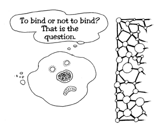
To bind or not to bind?
The ability of living cells to bind to each other or to the extracellular matrix between them is the foundation of the development of multicelled living organisms. It influences growth processes, changes in form, differentiation, nourishment, mobility, and cell death. Understanding the way a cell makes decisions may, therefore, advance our knowledge of the most elementary life processes, and should eventually lead to medical and industrial development.
Weizmann Institute scientists discovered that living cells can differentiate between crystals made of molecules that have exactly the same chemical composition, but whose structures are mirror images of each other. A cell appears to be able to choose between the surfaces of crystals of "left-handed" and "right-handed" molecules.
The scientists found that cells recognize and bind to external surfaces in one of two main ways: either quickly and strongly, or slowly and long-lived, i.e., the bond formed (with the surface or with another cell) exists for a long time. The scientists believe that for cells to examine a molecule surface or another cell, they have developed mechanisms that allow them to stay near the extracellular matrix, or close to each other, until they become more familiar with their surroundings and conditions are conducive to long-term connection. This is a short waiting and trial period, comparable to an engagement or cohabitation prior to a wedding. After this trial period, the cells are ready to develop steady and long-term contacts with the exterior.