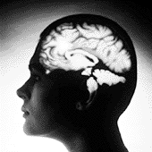
Basic research is clarifying the structure of the brain, its function, and its dysfunction. A communication receptor suspected of involvement in both Alzheimer’s disease and amyotrophic lateral sclerosis (ALS or Lou Gehrig’s disease) has been isolated at the Institute. In the future, an effective treatment for spinal and optic nerve injuries may ultimately result from successful experiments in nerve regeneration.
Institute scientists have developed new brain-imaging techniques that for the first time have made it possible to visualize the activity of millions of interconnected groups of neurons, enabling researchers to literally observe brain function with unprecedented resolution in time and space.
Discoveries revealing how the brain stores memories of taste and flavor may one day help overcome appetite disorders and lead to the development of medications to strengthen memory.
The Brain and the Central Nervous System
- Taste plus smell equals flavor
- A taste to remember
- Brain cells "tune in" to learn
- Nerve talk: too much can be deadly
- Pictures that tell important stories
- Repairing damage to the brain and spinal cord
- Teaching computers to see
- How the brain recognizes the images we see
- Why the brain likes smooth movements
The Brain and the Central Nervous System
Taste plus smell equals flavor

Weizmann Institute scientists investigated the way in which taste and smell sensations are stored in the memory. Information on taste is organized in the brain separately from that of smell; however, when the brain processes this information, nerve signals from the two senses unite and create a third, different representation. The latter represents flavor- the combination of taste and smell.
In collaboration with scientists from Washington, D.C., the researchers presumed that flavor is handled in a distinct region of the brain, separate from those where smell and taste are processed. They went on to demonstrate that they were right by discovering where taste and smell signals unite in the brains of rats.
This finding may serve as a springboard for future studies in learning and memory as well as studies on disorders of the taste and smell senses and problems such as loss of appetite.
A taste to remember
The molecular process that acts as a switch to activate the long-term memory of taste has been deciphered by Weizmann Institute researchers. The study focused on the region in the brain where the different taste memories are stored. Researchers discovered that taste sensations transmitted to the brain result in neural activity in this region, producing a defined change at the molecular level. This complex process, based on binding phosphorus atoms to specific proteins in the membrane of nerve cell terminals, aids in the long-term memory of different tastes.
Understanding these molecular changes may lead to the development of methodologies that will help in strengthening or weakening memory in general, and not only that related to taste.
Brain cells "tune in" to learn
Weizmann Institute scientists have proposed a new concept for understanding the cell mechanisms in the brain that are responsible for learning. This concept may shed new light on memory processes and eventually on aging and degenerative brain diseases.
The theory emanates from a new technology that allows researchers to meticulously examine the communication between two nerve cells in the brain. The technology revealed that earlier findings- that the learning process is expressed by an increase in the strength of the electric impulse flowing from the transmitting cell to the receptor cell- only told part of the story. There is actually a more complicated change in the character of the message transferred. The changes allow the connection to transmit information better or worse in specific bandwidths of frequencies of the stimulus. The transferred electrical information appears to be "fine tuned," rather like tuning a radio to a specific station.
The original study by Weizmann Institute researchers suggested that this fine and precise tuning for defined messages allows the brain cells to learn to manage a sort of "selective communication." The receiving cell "hears" or absorbs only the messages appropriate to it- that is, the messages to which it is "attuned." In a certain sense, the "learner" nerve cells behave like people trying to carry on a conversation in a noisy hall: they simply disregard the surrounding noise.
Nerve talk: too much can be deadly
Weizmann Institute scientists isolated one of the protein receptors of the neurotransmitter glutamate- the most common neurotransmitter stimulus in the brain- and later identified and isolated the gene responsible for the receptor's production.
More nerve communication junctions (known as synapses) secrete and absorb glutamate than any other neurotransmitter. Glutamate is involved, among other things, in memory and learning activities. However, when it is secreted in excessively large amounts, an exaggerated stimulus is created, expressed as an epileptic fit. It can also kill brain cells, for example, after a stroke or as a result of degenerative brain diseases such as Alzheimer's disease and Lou Gehrig's disease. Therefore, understanding the factors influencing the quantity of glutamate secreted in the brain and its mode of operation may produce new treatments for degenerative diseases, as well as ways of improving learning and memory processes.
The receptor isolated by Weizmann scientists acts like a camera shutter. When the neurotransmitter binds to it, it opens a channel through which ions flow, based on the difference in salt concentrations between the cell and its surroundings. Among other things, the newly-identified receptor may serve as a detector in the pharmaceutical industry.
Pictures that tell important stories
Scientists from the Weizmann Institute, Rockefeller University and IBM, with a background in a wide variety of research fields such as organic chemistry, spectroscopy, biophysics and the computer and optical sciences, innovated a methodology for examining the functional organization and timing of activities in the central nervous system.
This research method involves the use of optical imaging and novel dyes, sensitive to the electrical changes that occur in the nerve cell's membrane when an impulse is transferred across it. Sensitive cameras document this change, enabling researchers to examine the activity of millions of nerve cells simultaneously, in real time. Optical imaging has also been used to monitor changes in the color of the blood pigment hemoglobin when oxygen is transferred from red blood cells to nerve cells in the brain. (Oxygen is used by brain cells for, among other things, generating the energy needed for their electrochemical communication.) This real time monitoring of color-dependent activity changes allows scientists to study the spatial organization and activity of the various functional units in the brain.
Applications of these innovative research methods include researching how the brain organizes the electrical impulses it receives from the eye to form a significant image. It became clear from these studies that this organization is influenced by internal activity in the brain associated with different contexts. This means that two people can look at the same picture or listen to the same words and yet comprehend and develop entirely different sensations in reaction to it.
A medical use for the optical imaging system has recently been developed. This helps surgeons to locate functional borders during brain surgery to remove malignant tumors, and should reduce the risk of damage to healthy brain tissue.

Repairing damage to the brain and spinal cord
For many years it was thought that damage to the central nervous system (the brain and spinal cord) of higher animal species, including humans, could not be repaired. Injury to this system, for example, to the spinal cord, often results in irreversible paralysis. In contrast, lower animal species, such as fish and amphibians, can renew damaged nerve fibers and recover any function lost as a result of injury.
Weizmann Institute researchers, after examining the differences between lower and higher animals, suggested that evolutionary processes related to the interaction between the central nervous system and the immune system may have ended the ability to regenerate nerve fibers in the central nervous system. This new concept raises hopes for developing innovative methods, involving treatment by the body's own immune cells, to achieve regeneration of injured nerve fibers in the central nervous system. In the future, these cell therapies may be used to treat people with injuries to the central nervous system, particularly in the spinal cord.
Vision and Robotics
Teaching computers to see
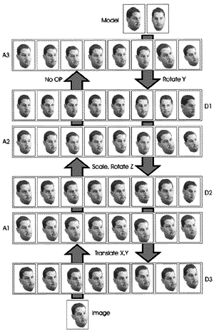
Mathematicians at the Weizmann Institute have designed ways to enable computerized vision systems to recognize objects by using a limited quantity of photographs taken from a variety of angles. These computerized "eyes" should enable robots to move and find their way along changing paths in production facilities, as well as assist them in identifying objects. The scientists involved in this area are collaborating with the Institute's brain researchers, whose work indicates that the human brain identifies objects in ways similar to those vision systems developed by the mathematicians.
How the brain recognizes the images we see

It was previously thought that when the brain receives new visual data, it locates it initially in the primary vision area and then transfers it to the higher vision area. However, it has become evident that the transfer path also works in the opposite direction: from the higher vision area to the primary vision area.
Weizmann Institute mathematicians have developed a functional model to explain this discovery. The specific visual data that is sent from the higher to the lower vision area is determined by which new visual data is initially sent from the lower to the higher vision area, i.e., the things that we are seeing at any one moment. In this way, the two streams of visual data flow past each other at very high speeds, enabling the brain to compare large numbers of pictures and to quickly identify any similarities between them.
This proposed model is of great importance in brain research and in the development of robot applications, both of which are based on imitating the human brain's functioning and abilities.
Why the brain likes smooth movements
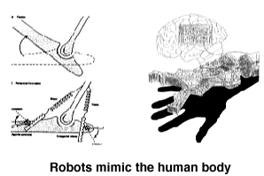
Scientists from the Weizmann Institute have developed a mathematical model that describes how the human brain plans and monitors the movement of the body's limbs. The model is based on the idea that the desired movements are represented within the brain in terms of spatial coordinates; the desired movements are then executed by appropriate activation of the nerves and muscles. This model uses an original theory developed by Institute researchers in collaboration with Massachusetts Institute of Technology colleagues. The model states that the primary factor in how the brain plans certain movements of the upper limbs, such as extending the arm, is that it should be executed smoothly. This concept relates to the rate at which the acceleration of the arm changes, as expressed in spatial coordinates.
By analyzing the movement precisely, a very detailed prediction can be made of the trajectories and characteristics of motion that a person will carry out. In other experiments, a precise analysis of movement is used to examine the differences between how healthy people move their limbs, and how the same motions are performed by people suffering from degenerative brain diseases, such as Parkinson's disease. Examining these differences helps to understand the link between damage to a particular area of the brain that is known to be affected by Parkinson's disease, and failure in one or several of the components used in planning and executing the movement.
The mathematicians are collaborating with researchers from other fields, including brain research, with the goal of discovering and understanding the language that the brain uses to plan and organize the body's movement. The results are also useful in robotics research in developing strategies that can allow robot movements to be planned and carried out efficiently.


